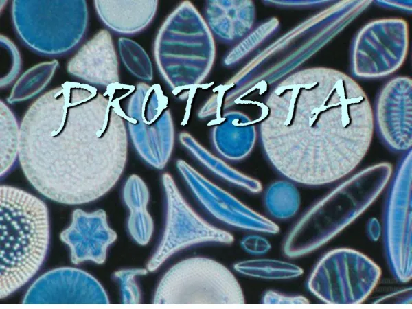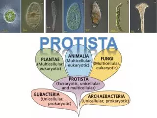
Structural and Functional Diversity of Protists in Eukaryotic Evolution
Explore the diverse world of protists, the most nutritionally diverse group of eukaryotes. Learn about their structural complexity, modes of nutrition, and the role of endosymbiosis in their evolution. Discover fascinating protist groups like ciliates, sarcodines, forams, radiolarians, apicomplexans, zooflagellates, euglenophytes, and dinoflagellates. Understand their characteristics, behaviors, and their impact on human health and the environment.


Structural and Functional Diversity of Protists in Eukaryotic Evolution
E N D
Presentation Transcript
Fig. 28-03g 5 µm
Fig. 28-03h 50 µm
Fig. 28-06 9 µm
Fig. 28-03j 20 µm 50 µm
Fig. 28-04 Flagella Undulating membrane 5 µm
Most eukaryotes are single-celled organisms • Protists are eukaryotes and thus have organelles and are more complex than prokaryotes • Most protists are unicellular, but there are some colonial and multicellular species (ex. kelp)
Structural and Functional Diversity in Protists • Protists exhibit more structural and functional diversity than any other group of eukaryotes • Single-celled protists can be very complex, as all biological functions are carried out by organelles in each individual cell
Protists, the most nutritionally diverse of all eukaryotes, include: • Photoautotrophs, which contain chloroplasts • Heterotrophs, which absorb organic molecules or ingest larger food particles • Mixotrophs, which combine photosynthesis and heterotrophic nutrition
Endosymbiosis in Eukaryotic Evolution • There is now considerable evidence that much protist diversity has its origins in endosymbiosis • Mitochondria evolved by endosymbiosis of an aerobic prokaryote • Plastids evolved by endosymbiosis of a photosynthetic cyanobacterium
Theory of Endosymbiosis • Animation - Endosymbiosis
Classification- based on method of obtaining nutrition • Animal-like- protozoans- consume other organisms, some parasites • Plant-like- algae- make their own food, some can consume other organisms when light is unavailable • Fungus-like- feed on decaying organic matter and absorb nutrients through cell walls, some can consume others, few are parasites
Ciliates • Ciliates, a large varied group of protists, are named for their use of cilia to move and feed • They have large macronuclei and small micronuclei • The micronuclei function during conjugation, a sexual process that produces genetic variation • Conjugation is separate from reproduction, which generally occurs by binary fission
Phylum Ciliophora – move by cilia i.e. Parmecium • Ciliate Movement • Paramecium Feeding • Paramecium Conjugation
Fig. 28-11a Contractile vacuole Oral groove Cell mouth Cilia 50 µm Micronucleus Food vacuoles Macronucleus (a) Feeding, waste removal, and water balance
Sarcodina • Amoebas move and feed by pseudopodia • http://www.cbsnews.com/8301-204_162-57528956/brain-eating-amoeba-kills-10-in-pakistan-how-rare-is-it/
Fig. 28-03l 100 µm
Phylum Sarcodina – animals move and feed by pseudopodia/cytoplasmic streamingFeeding - Animation – Cytoplasmic Streaming
Forams • Foraminiferans, or forams, are named for porous, generally multichambered shells, called tests • Pseudopodia extend through the pores in the test • Foram tests in marine sediments form an extensive fossil record
Fig. 28-03i 20 µm
Radiolarians • Marine protists called radiolarians have tests fused into one delicate piece, usually made of silica • Radiolarians use their pseudopodia to engulf microorganisms through phagocytosis • The pseudopodia of radiolarians radiate from the central body
Fig. 28-18 Pseudopodia 200 µm
Apicomplexans • Apicomplexans are parasites of animals, and some cause serious human diseases • One end, the apex, contains a complex of organelles specialized for penetrating a host • Most have sexual and asexual stages that require two or more different host species for completion
The apicomplexan Plasmodium is the parasite that causes malaria • Plasmodium requires both mosquitoes and humans to complete its life cycle • Approximately 2 million people die each year from malaria • Efforts are ongoing to develop vaccines that target this pathogen
Fig. 28-10-3 Inside mosquito Inside human Merozoite Sporozoites (n) Liver Liver cell Oocyst Apex MEIOSIS Red blood cell 0.5 µm Merozoite (n) Zygote (2n) Red blood cells FERTILIZATION Gametes Key Gametocytes (n) Haploid (n) Diploid (2n)
Zoomastigina • Zooflagellates • Move using flagella • Some are parasitic, others feed on other organisms • Cause diseases such as American Sleeping Sickness (Chagas) and African Sleeping Sickness
Euglenophyta • Euglena have one or two flagella that emerge from a pocket at one end of the cell • Some species can be both autotrophic and heterotrophic
Fig. 28-07 Long flagellum Eyespot Short flagellum Light detector Contractile vacuole Nucleus Chloroplast Plasma membrane Pellicle Euglena (LM) 5 µm
Dinoflagellates • Dinoflagellates are a diverse group of aquatic mixotrophs and heterotrophs • They are abundant components of both marine and freshwater phytoplankton • Each has a characteristic shape that in many species is reinforced by internal plates of cellulose
Two flagella make them spin as they move through the water • Dinoflagellate blooms are the cause of toxic “red tides” http://www.whoi.edu/redtide/
Fig. 28-09 Flagella 3 µm
Diatoms • Diatoms are unicellular algae with a unique two-part, glass-like wall of hydrated silica • Diatoms usually reproduce asexually, and occasionally sexually
Fig. 28-13 3 µm
Diatoms are a major component of phytoplankton and are highly diverse • Fossilized diatom walls compose much of the sediments known as diatomaceous earth
Golden Algae (Chrysophyta) • Golden algae are named for their color, which results from their yellow and brown carotenoids • The cells of golden algae are typically biflagellated, with both flagella near one end • All golden algae are photosynthetic, and some are also heterotrophic • Most are unicellular, but some are colonial
Fig. 28-14 Flagellum Outer container Living cell 25 µm
Brown Algae (Phaeophyta) • Brown algae are the largest and most complex algae • All are multicellular, and most are marine • Brown algae include many species commonly called “seaweeds” • Brown algae have the most complex multicellular anatomy of all algae
Giant seaweeds called kelps live in deep parts of the ocean • The algal body is plantlike but lacks true roots, stems, and leaves and is called a thallus • The rootlike holdfast anchors the stemlike stipe, which in turn supports the leaflike blades
Fig. 28-15 Blade Stipe Holdfast
Red Algae (Rhodophyta) • Red algae are reddish in color due to an accessory pigment call phycoerythrin, which masks the green of chlorophyll • The color varies from greenish-red in shallow water to dark red or almost black in deep water • Red algae are usually multicellular; the largest are seaweeds • Red algae are the most abundant large algae in coastal waters of the tropics
Fig. 28-19 Bonnemaisonia hamifera 20 cm 8 mm Dulse (Palmaria palmata) Nori. The red alga Porphyra is the source of a traditional Japanese food. The seaweed is grown on nets in shallow coastal waters. The harvested seaweed is spread on bamboo screens to dry. Paper-thin, glossy sheets of nori make a mineral-rich wrap for rice, seafood, and vegetables in sushi.
Green Algae (Chlorophyta) • Green algae are named for their grass-green chloroplasts • Plants are descended from the green algae • The two main groups are chlorophytes and charophyceans
Most chlorophytes live in fresh water, although many are marine • Other chlorophytes live in damp soil, as symbionts in lichens, or in snow
Chlorophytes include unicellular, colonial, and multicellular forms
Fig. 28-21 (a) Ulva, or sea lettuce 2 cm (b) Caulerpa, an intertidal chloro- phyte
Fig. 28-21b (b) Caulerpa, an intertidal chloro- phyte
Oomycetes (Water Molds , etc.) • Oomycetesinclude water molds, white rusts, and downy mildews • They were once considered fungi based on morphological studies • Most oomycetes are decomposers or parasites • They have filaments (hyphae) that facilitate nutrient uptake • Their ecological impact can be great, as in Phytophthorainfestans causing potato blight