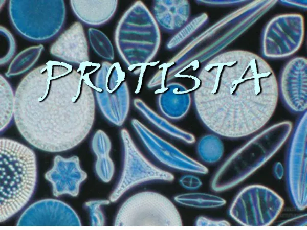
Protista
Protista. Slideshow By: Paola Saavedra Anna Iszard. Diplomonadia. Structure: Diplomonadia contain multiple flagella, two separate nuclei, a relatively simple cytoskeleton, no plastids, and no mitochondria. Interaction with humans:

Protista
E N D
Presentation Transcript
Protista Slideshow By: Paola Saavedra Anna Iszard
Diplomonadia • Structure: • Diplomonadia contain multiple flagella, two separate nuclei, a relatively simple cytoskeleton, no plastids, and no mitochondria. • Interaction with humans: • Diplomonadia is a parasite that infects the human intestine, causing abdominal cramps and severe diarrhea to occur. • Someone can pick up Giardia by drinking water that is contaminated with human feces containing the parasite in a dormant cyst stage. • Uniqueness: • Boiling drinking water that contains diplomonadia kills the cysts. • Example: • Giardialamblia
Parabasalida • Structure: • Parabasalida lack mitochondria and contain flagella. • Interaction with humans: • This is a common inhabitant of the vagina of human females. • It can breed rapidly and infect the vaginal lining by overcoming beneficial microbial populations if the normal acidity of the vagina is disturbed. • These infections can also occur in the urethras of males, though often without symptoms. • Sexual transmission can spread the infection. • Uniqueness: • T. vaginalis has both flagella and an undulating membrane, which helps it move along the mucus-coated membrane within the reproductive and urinary tracts of their human hosts. • Example: • Trichomonasvaginalis
The Euglenophyta group is separated into two main categories: Euglenoids and Kinetoplastids
Euglenoids • Structure: • Euglenoids are known for having an anterior pocket, or chamber, from which one or two flagella emerge. • Interaction with humans: • They fight off germs and pollutants in our pond water. • Uniqueness: • Euglenoids are unicellular and are autotrophic. • Contains Paramylon, a glucose polymer that functions as a storage molecule. • Example: • Euglena
Kinetoplastids • Structure: • Kinetoplastids house a single large mitochondrion associated with a kinetoplast, which houses extranuclear DNA. • Interaction with humans: • A species of Trypanosoma, a type of Kinetoplastid, causes an African sleeping sickness disease that is spread by the bite of the Tsetse fly. • Uniqueness: • Kinetoplastids are symbiotic, and some are pathogenic to their hosts. • Example: • Trypanosoma
The Alveolata group is separated into three main categories: Apicomplexans, ciliates, and Dinoflagellates.
Apicomplexans • Structure: • Apicomplexans are groups of parasites. • Interaction with humans: • Can cause serious diseases in humans, like Malaria. • Uniqueness: • All apicomplexans are parasites of animals. • The parasties spread as tiny infectious cells called sporozoites. • Reproduction: • Apicomplexans have sexual and asexual stages throughout its life, and these cycles often require two or more different host species for completion. • Example: • Plasmodium
Ciliates • Structure: • Ciliates use their cilia for movement and feeding. • These cilia are shorter than flagella. • Interaction with humans: • Ciliates do not interact with humans. • Uniqueness: • Ciliates live as solitary cells in fresh water. • In ciliates, there is a presence of two types of nuclei, a large macronucleus and usually several tiny micronuclei. • Reproduction: • Ciliates reproduce through binary fission, where the macronucleus extends and splits, rather than undergoing mitotic division. • Example: • Stentor
Dinoflagellates • Structure: • Most dinoflagellates are unicellular, but some are colonial forms. • Dinoflagellate have a characteristic shape which is reinforced in some species by internal plates of cellulose. • The movement of two flagella in perpendicular grooves in this cellulose produces a spinning movement for which these organisms are named. • Interaction with humans: • Fish deaths have increased in coastal waters of the U.S. mid-Atlantic states in the past decade, possibly a result of pollution of the water with fertilizers containing dinoflagellates, affecting food supply for humans. • Dinoflagellates give off light, creating an eerie ocean glow at night when waves, boats, or human swimmers agitate water containing dense populations of the dinoflagellates.
Dinoflagellates Cont. • Uniqueness: • Dinoflagellate blooms(episodes of explosive population growth) cause red tides in coastal waters. • During these blooms, its toxin stuns fish, and the dinoflagellates feed on their prey’s body fluids. • Some dinoflagellates are carnivorous. • Some dinoflagellates are bioluminescent(glow). • When the surrounding water is agitated by small predators that feed on phytoplankton, the light emitted by the dinoflagellates attracts fish that eat those smaller predators. • Some dinoflagellates live as mutualisticsymbionts of animals called cnidarians that build coral reefs, where the photosynthetic output of the dinoflagellates is the main source of food for the reef community. • Example: • Pfiesteriapiscicida(on previous slide) • Noctilucales(on current slide)
References • Photos: • Carr, J. (2006). “Giardialamblia SEM 8698 lores”. [Photograph]. Retrieved February 15, 2012, from The Centers for Disease Control and Prevention: http://phil.cdc.gov/PHIL_Images/8698/8698_lores.jpg • Colm, G. (2010). “Trichomonasvaginalis phase contrast microscopy”. [Photograph]. Retrieved February 21, 2012, from Wikipedia: http://upload.wikimedia.org/wikipedia/commons/2/27/Trichomonas_vaginalis_phase_contrast_microscopy.jpg • Miklos, C. (2011). “Euglena diagram”. [Photograph]. Retrieved February 15, 2012, from Simple English Wikipedia: http://en.wikipedia.org/wiki/File:Euglena_diagram.jpg • Schultz, M. (1970). “Trypanosoma sp. PHIL 613 lores”. [Photograph]. Retrieved February 15, 2012, from The Centers for Disease Control and Prevention: http://phil.cdc.gov/phil/details.asp?pid=613 • Hollings Marine Laboratory. (2007). “Pfiesteriapiscicida”. [Photograph]. Retrieved February 21, 2012, from Science Daily: http://images.sciencedaily.com/2007/01/070119164427.jpg • PLoS Biology. (2005). “Malaria”. [Photograph]. Retrieved February 21, 2012, from Wikipedia: http://upload.wikimedia.org/wikipedia/commons/f/f1/Malaria.jpg • Quadell. (2005). “Stentorroeseli composite image”. [Photograph]. Retrieved February 21, 2012, from Wikipedia: http://upload.wikimedia.org/wikipedia/commons/5/5a/Stentor_roeseli_composite_image.jpg • Nilsson, J. A. (2001). “Volvox”. [Photograph]. Retrieved February 21, 2012, from Sharon Taxonomy: http://sharon-taxonomy2009-p3.wikispaces.com/Protista • Sampayo, M. A. (2007). “Noctilucascintillansunica”. [Photograph]. Retrieved February 21, 2012, from Wikipedia: http://upload.wikimedia.org/wikipedia/commons/6/62/Noctiluca_scintillans_unica.jpg
References Cont. • Text: • Campbell, N. A., & Reece, J. B. (2002). Biology. (6th ed. ed.). New York: Benjamin Cummings. • Edwards, G. I., & Pfirrmann, C. (2010). E-z biology. Barrons Educational Series Inc. • The Princeton Review. (2011). Cracking the sat biology e/m subject test. Princeton, New Jersey: The Princeton Review. • Aldridge, C., & Croston, G. E. (2011). Sat subject test: Biology e/m 2011-2012. New York, NY: Kaplan Publishing.