
Apoptosis (Programmed Cell Death)
E N D
Presentation Transcript
Apoptosis vs Necrosis • Level of stress, change in environment stress apoptosis necrosis
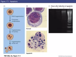
A pathological response to injury Chromatin clumping Mitochondria swelling and rupture Plasma membrane lyses Cell contents spill out A normal physiological response to specific suicide signals Chromatin condenses Internucleosomal cleavage leads to laddering of DNA Cytoplasma shrinks without membrane rupture Blebbing of plasma and nuclear membranes No spillage Modes of Cell Death Necrosis Apoptosis
Tumor Suppressor Genes • Tumor-suppressor genes, function like brakes, keep cell numbers down, either by: 1. Inhibiting progress through the cell cycle and thereby preventing cell birth, or 2. Promoting apoptosis • When cellular tumor suppressor genes are rendered non-functional through mutation, the cell becomes malignant. Example: pRb, p53, and p16INK4a.
Oncogenes • Oncogenes stimulate cells to grow (or refuse to die). • Mutated oncogenes can stimulate cells even when they are not receivinggrowth signals. Example: Ras, Akt, Survivin. • Amplification of oncogenes is also found in cancer. Example: MDM2.
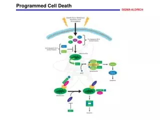
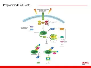
Apoptosis Signaling Pathway Cell, 2002;108:153-64
Role of p53 in Apoptosis • p53 induces apoptosis through transcriptional activation of proapoptotic genes, such as Puma, Noxa, p53AIP, Bax, Apaf-1, etc. • It can also directly induce apoptosis by localizing to mitochondria via interaction with Bcl-2 family protein. Nat Rev Cancer, 2002;2:594-604
Tumor Suppressor p53 • Mutated/inactivated in a majority of human cancers • p53 -/- knockout mice embryos develop normally to term but after a few weeks the young mice develop numerous malignant tumors • adenovirus introduced wild-type p53 markedly enhanced the anti-tumor effect of a common chemotherapeutic agent, cisplatin, in human non-small cell lung cancer cells, and in human colon cancer cells. • Effectiveness of cancer therapy correlates with the ability to induce p53-dependent apoptotic response