Download
1 / 45
450 likes | 470 Views
Learn the basics of fMRI data modeling and statistical inference. Explore signal modeling, noise statistics, spatial and temporal activation models, and more. Discover the challenges and nuances of analyzing fMRI data and evaluating different methods. Dive into the principles underlying most fMRI analyses, especially the General Linear Model. Gain insights into the Hemodynamic Response Function, activation blobs, linear convolution, and variable shape HRF analysis.

E N D
FMRI Data Modeling,the General Linear Model,and Statistical Inference Robert W Cox, PhD SSCC/NIMH/NIH/DHHS/USA/EARTH http://afni.nimh.nih.gov/pub/tmp/ISMRM2007/ fMRI: Basics to Cutting Edge – ISMRM 2007 – Berlin – 19 May 2007
Assumptions about You • You sort-of-know a little about how FMRI works • e.g., You’ve paid attention today? • You want to sort-of-know a little about mathematics of FMRI analysis • So you can read papers? • So you can judge how appropriate an analysis method is for your work? • So you can start hacking out code?
Caveats • Almost everything herein has an exception or complication, or both • Special types of data or stimuli may require special analysis steps • e.g., perfusion-weighted FMRI • Special types of questions often require special data and analyses • e.g., relative timing of neural events
Outline • Signal Modeling Principles • e.g., generic ranting • Temporal Models of Activation • e.g., convolution • Noise Models & Statistics • e.g., prewhitening, resampling • Spatial Models of Activation • e.g., clustering, smoothing, ROIs
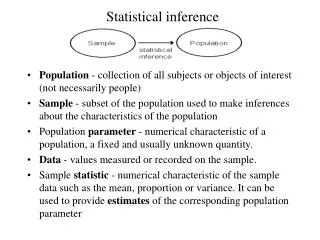
Signal Modeling Principles • Develop a mathematical model relating what we know(stimulus timing and image data) to what we want to know(location, amount, timing, etc, of neural activity) • Given data, use this model to solve for unknown parameters in the neural activity (e.g., when, where, how much, etc) • Then test for statistical significance
The Data • 10,000..50,000 image voxels inside brain (resolution 2-3 mm) • 100..1000+ time points in each voxel (time step 2 s) • Also know timing of stimuli delivered to subject (etc) • Behavioral, physiological data? • Hopefully, some hypothesis
One slice at one time; Blue box shows graphed voxels Graphs of 33 voxels through time Sample Data: Visual Area V1
Same Data as Last Slide This is reallygood data; N.B.: repetitions differ Blowup of central time series graph: about 7% signal change with a very powerful periodic neural stimulus Block design experimental paradigm: visual stimulation
Event-Related Data Four different visual stimuli • White curve = Data (first 136 TRs) • Orange curve = Model fit (R2=50%) • Green = Stimulus timing Very good fit for ER data (R2=10-20% more usual). Noise is as big as BOLD!
Why FMRI Analysis Is Hard • Don’t know true relation between neural “activity” and BOLD signal: • What is neural “activity”, anyway? • What is connection between “activity” and hemodynamics and MRI signal? • Noise in data is poorly characterized • In space and in time, and in origin • Noise amplitude BOLD signal • Can some of this noise be removed? • Makes both signal detection and statistical assessment hard
Why So Many Methods? • Different assumptions about activity-to-MRI signal connection • Different assumptions about noise (signal fluctuations of no interest) properties and statistics • Different experiments and questions • Result: Many “reasonable” FMRI analysis methods • Researchers must understand the tools!! (Models and software)
Fundamental Principles Underlying Most FMRI Analyses (esp. GLM):HRF Blobs • Hemodynamic Response Function • Convolution model for temporal relation between stimulus and response • Activation Blobs • Contiguous spatial regions whose voxel time series fit HRF model • e.g., Reject isolated voxels even if HRF model fit is good there
Temporal Models:Linear Convolution • Additivity Assumption: • Input = 2 separated-in-time activations • Output = separated-in-time sum of 2 copies of the 1-stimulus response • FMRI response to single stimulus is called the Hemodynamic Response Function (HRF) • Also: Impulse Response Function (IRF)
Brief Stimulus at time t = 1 Model function h(t) = t8.6e–t/0.547 (Mark Cohen) Simple Model HRF
“Event-Related” Stimuli at times t = 1,7,10 Signal = HRF Stimulus
Ideal response to 1 brief stimulus 220 sec stimulus blocks Block Stimulus
Some (incomplete) Signal Models • One stimulus class: stimuli occur at times s HRF: the analysis target! • One stimulus class: stimulus/activity occurs in 2 separated phases Stimulus time • Models must be adjusted to particular experimental design Delay between phases
Fixed Shape HRF Analysis • Assume some shape for HRF=h(t) • Signal model is r(t) =h(t)Stimulus = “Convolution” of HRF with neural activity timing function (e.g., stimulus) • Model for each voxel data time series: Z(t) = ar(t) + b + noise(t) • Estimate unknowns: a=amplitude, b=baseline, 2 =noise variance • Significance of a≠0activation map
Variable Shape HRF Analysis • Allow shape of HRF to be unknown, as well as amplitude (deconvolution) • Good: Analysis adapts to each subject and each voxel • Good: Can compare brain regions based on HRF shapes • e.g., early vs. late response? • Bad: Must estimate more parameters • Need more data (all else being equal)
Aside: Baseline Model • Need to model a slowly drifting baseline, since the signal from people fluctuates on time scale of 100 s or so • Mostly due to tiny movements? • Scanner fluctuations can also occur • Usual method: include low frequency expansion in signal model (“highpass filtering”):
HRF Model Equations Simplest model: fixed shape Unknown = a[b&cfixed] Next simplest model: derivative allows for time shift Unknowns = a0anda1 [b&cfixed] Expansion in a set of fixed basis functions {q(t)} (e.g., Splines, sines, …); Unknowns = {wq}
Multiple Stimulus Classes • Need to calculate HRF (amplitude or amplitude+shape) separately for each class of stimulus • Novice FMRI researcher pitfall: try to use too many stimulus classes • Event-related FMRI: need 20+ events per stimulus class • Block design FMRI: need 10+ blocks per stimulus class
Combined Signal Model Convolution HRF model Reorder sums • Result: equation for unknowns {0, 1, wq} in terms of data Z(t)
Matrix-Vector Formulation • Usually write equation in form: • In matrix-vector notation: Each column of Ris a time series basis function, and each element of is its amplitude in z
Sample Variable HRF Analysis ‘What’ HRF ‘Where’ HRF Where HRF What HRF • ‘What’-vs-‘Where’ tactile stimulation • Red regions with What Where Data from R van Boven: 1040 time points; 30 stimuli in each class
(Linear) Inverse Modeling • Instead of using stimulus timing to get HRF, could use an assumed HRF to get activity timing per voxel • Or could use an assumed spatial response (from a training/calibration run?) to extract stimulus timing • e.g., HBM 2006 Movie contest • Linear equations, buthave swapped roles of unknowns & knowns
Noise Models & Statistics • Physiological “noise” • Heartbeat and respiration affect signal in complex ways • Subject head movement • After realignment, some effects remain • Low frequency drifts ( 0.01 Hz) • Scanner glitches can produce gigantic (10 ) spikes in data
Physiological “Noise” • MRI signal changes due to non-neural physiology during scan • Can be approximately filtered out with external measurements • e.g., respiratory bellows, pulse oximeter • Somewhat harder than it sounds, and is not commonly used (yet)
Fluctuations: 16 images/sec (one slice) 0.22 Hz 1.08 Hz
Regression Methods • Solving this equation approximately: • What method to use to solve for ? • Can allow for statistics of in solution method • Should allow for statistics of in solution statistics • Neither of these points are trivial, fully-resolved issues Ris NxM matrix z&are N-vectors is M-vector (M<<N)
Regression Methods I • Ordinary least squares: • Derivable under assumption that has N(0,2I) distribution (Gaussian white noise) • Pro: simple, standard, robust • Con: not as statistically powerful as possible • Prewhitened least sqrs: • Derivable under assumption that has N(0,C) distribution (C = covariance matrix) • Pro: as statistically powerful as possible given the assumptions • Con: sensitive to estimation of C
Regression Methods II • Projected least squares: • P = projection matrix, onto “acceptable” subspace of data • Pro: can remove à priori unwanted components from data (e.g., low and high frequencies) • L1 regression: • Pro: robust against non-Gaussianity in • Con: harder to estimate significance of analytically; temporal correlation is also harder to handle
Inference on • contains the results about the HRF • Can test individual elements in or collections of elements for significant difference from zero (“activation”) • e.g., “was there a response to stimulus A?” • Can test differences between elements or collections of elements • e.g., “was response to A different from B?” • Tests usually expressed as t or F statistic
Estimating Serial Correlation • Can assume some model correlation structure; e.g., AR(n) autoregressive models • Advantage is simplicity, not reality • Can try to estimate C directly • Possibly using neighboring voxels as well • Or smooth estimates of C (or some of the parameters in C) locally • Usually start with OLS to estimate and subtract “signal”, then estimate C from residuals
Adapting to Correlated Noise • Can adjust degrees-of-freedom in OLS estimates of parameters to approximate for correlation • Including correlation induced by projection via bandpass filters • If “properly” done, prewhitened LS will give full degrees-of-freedom with no semi-ad hoc adjustments required • Results can be sensitive to errors in C
Avoiding Some Assumptions • All statistical methods require assumptions about noise • Gaussianity, independence, … • Can use modern statistical resampling/permutation methods to reduce the number of assumptions • Very computationally intensive • Substituting number crunching for mathematical theory
Spatial Models of Activation • 10,000..50,000 image voxels in brain • Don’t really expect activation in a single voxel (usually) • Curse of multiple comparisons: • If have 10,000 statistical tests to perform, and 5% give false positive, would have 500 voxels “activated” by pure noise — way way too much! • Can group voxels together somehow to manage this curse
Spatial Grouping Methods • Smooth data in space before analysis • Average data across anatomically-selected regions of interest ROI (before or after analysis) • Labor intensive (i.e., send more postdocs) • Reject isolated small clusters of above-threshold voxels after analysis
Spatial Smoothing of Data } Good things • Reduces number of comparisons • Reduces noise (by averaging) • Reduces spatial resolution • Can make FMRI results look PET-ish • In that case, why bother gathering high resolution MR images? • Smart smoothing: average only over nearby brain or gray matter voxels • Uses resolution of FMRI cleverly • Or: average over selected ROIs • Or: cortical surface based smoothing
Spatial Clustering • Analyze data, create statistical map (e.g., t statistic in each voxel) • Threshold map at a lowish tvalue, in each voxel separately • Threshold map by rejecting clusters of voxels below a given size • Can control false-positive rate by adjusting t threshold and cluster-size thresholds together
What the World Needs Now • Unified HRF/Deconvolution⊕Blob analysis • Time⊕Space patterns computed all at once, instead of via arbitrary spatial smoothing • Increase statistical power by using data from multiple voxels cleverly • Instead of time analysis followed by spatial analysis (described earlier) • Instead of component-style analyses (e.g., ICA) that do not use stimulus timing or other known info • Must be grounded in realistic brain+signal models • Difficulty: models for spatial blobs • Little information à priori⇒ must be adaptive
Inter-Subject Analyses • Bring brains into alignment somehow • Perform statistical analysis on activation amplitudes • e.g., ANOVA of various flavors • Can be cast as a similar regression problem, with “data” = • Not yet tried much: analyze all subjects’ time series together at once in one humungous regression
Summary and Conclusion • FMRI data contain features that are about the same size as the BOLD signal and are poorly understood • Thus: There are many “reasonable” ways to analyze FMRI data • Depending on the assumptions about the brain, the signal, and the noise • Conclusions: Understand what you are doing & Look at your data • Or you will do something stupid
Finally … Thanks • The list of people I should thank is not quite endless … MM Klosek. JS Hyde. JR Binder. EA DeYoe. SM Rao. EA Stein. A Jesmanowicz. MS Beauchamp. BD Ward. KM Donahue. PA Bandettini. AS Bloom. T Ross. M Huerta. ZS Saad. K Ropella. B Knutson. J Bobholz. G Chen. RM Birn. J Ratke. PSF Bellgowan. J Frost. K Bove-Bettis. R Doucette. RC Reynolds. PP Christidis. LR Frank. R Desimone. L Ungerleider. KR Hammett. DS Cohen. DA Jacobson. EC Wong. D Glen. Et alii … http://afni.nimh.nih.gov/pub/tmp/ISMRM2007/