Download

1 / 18
230 likes | 701 Views
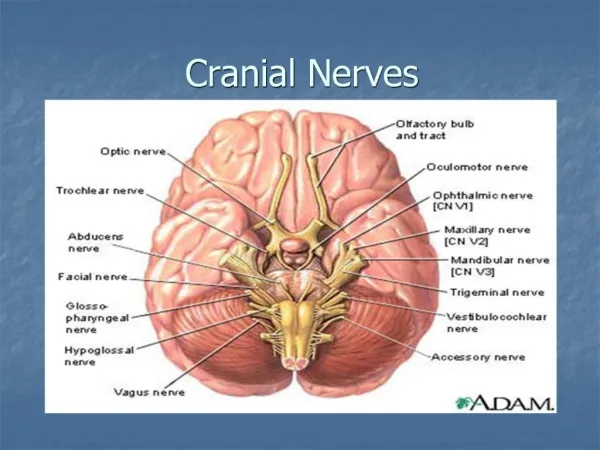
Cranial Neves IX, X & XI. Dr. Nimir Dr. Safaa. Glossopharyngeal Nerve (Cranial Nerve IX) It is a motor and a sensory nerve. Glossopharyngeal nerve n uclei are three nuclei: ( 1) M ain motor nucleus(SVE). ( 2) P arasympathetic nucleus(GVE). (3 ) S ensory nucleus(GVA).

E N D
CranialNeves IX, X & XI Dr. Nimir Dr. Safaa
Glossopharyngeal Nerve (Cranial Nerve IX) • It is a motor and a sensory nerve. • Glossopharyngeal nerve nuclei are three nuclei: • (1) Main motor nucleus(SVE). • (2) Parasympathetic nucleus(GVE). • (3) Sensory nucleus(GVA). • Main Motor Nucleus: • It lies deep in reticular formationof medulla oblongata and is formed by superior end of nucleus ambiguus. • It receives corticonuclear fibers from both cerebral hemispheres. • Efferentfibers supply stylopharyngeus muscle.
Parasympathetic Nucleus: • It is also called inferior salivatorynucleus. • It receives: • Afferentfibers from hypothalamus through descending autonomic pathways. • Information from olfactory system through reticular formation. • Information concerning taste from nucleus of solitary tract from mouth cavity. • Efferent preganglionic parasympathetic fibers reach otic ganglion through tympanicbranch of glossopharyngeal, tympanic plexus, and lesser petrosalnerve. • Postganglionicfibers pass to parotid salivary gland.
Sensory Nucleus: • It is part of nucleus of tractussolitarius. • Sensations of taste travel through peripheral axons of nerve cells situated in ganglionon glossopharyngeal nerve. • Central processes of these cells synapse on nerve cells in sensory nucleus. • Efferentfibers cross and ascend to ventral nuclei of thalamusand hypothalamicnuclei. • From thalamus through internal capsule and corona radiatafibers end in lower part of the postcentralgyrus.
Afferent concerns common sensation enters brainstem throughsuperior ganglion of glossopharyngealbut ends in spinal nucleus of trigeminal. • Afferentimpulses from carotid sinus baroreceptor travel with glossopharyngeal & terminate in nucleus of tractussolitariusand connected to dorsal motor nucleus of vagusnerve. • Carotid sinus reflex that involves glossopharyngeal and vagus nerves assists in regulation of arterial blood pressure.
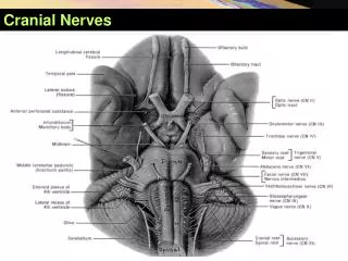
Course of Glossopharyngeal Nerve: • It leaves anterolateral surface of upper medullaoblongata between olive and inferior cerebellar peduncle. • It passes laterally in posterior cranial fossa and leavesskull through jugular foramen. • Superior and inferior glossopharyngeal sensory ganglia are situated on nerve here. • The nerve descends through upper part of the neck with internal jugular vein and internal carotid artery to reach posterior border of stylopharyngeusmuscle, which it supplies. • The nerve then passes forward between superior and middle constrictor muscles of pharynx to give sensory branches to mucous membrane of pharynx and posterior third of tongue.
Vagus Nerve (Cranial Nerve X) • It is a motor and a sensory nerve. • Vagus Nerve Nuclei: • It has three nuclei: • (1) Mainmotornucleus. • (2) Parasympathetic nucleus. • (3) Sensorynucleus. • Main Motor Nucleus: • It lies deep in reticular formation of medulla oblongata and is formed by nucleus ambiguus. • It receives corticonuclear fibers from both cerebral hemispheres. • The efferent fibers supply constrictor muscles of pharynx intrinsic muscles of the larynx.
Parasympathetic Nucleus: • It forms dorsal nucleus of vagus and lies beneath floor of lower part of fourth ventricle posterolateralto hypoglossal nucleus. • It receives: • Afferent fibers from hypothalamusthrough descending autonomic pathways. • Afferents from glossopharyngeal(carotid sinus reflex). • Efferentfibers are distributed to involuntary muscle of bronchi, heart, esophagus, stomach, small intestine, and large intestine as far as distal one-third of transverse colon.
Sensory Nucleus: • It is lower part of nucleus of tractussolitarius. • Sensations of taste travel through peripheral axons of nerve cells situated in inferior ganglion on vagus nerve. • Centralprocesses of those cells synapse on nerve cells in sensory nucleus. • Efferentfibers cross the median plane and ascend to ventral nuclei of thalamus & hypothalamic nuclei. • From thalamus through internal capsule and corona radiata fibers end in postcentralgyrus. • Afferent concerns common sensation enters brainstem through superior ganglion of vagusbut ends in spinal nucleus of trigeminal.
Course of the Vagus Nerve • It leaves anterolateralsurface of upper medulla oblongata between oliveand inferior cerebellar peduncle. • It passes laterally through posteriorcranial fossa and leaves skull through jugular foramen. • The vagus nerve has two sensory ganglia: • Rounded superior on the nerve within jugular foramen. • Cylindrical inferiorwhich lies on the nerve just below the foramen. • At inferior ganglion, cranial root of accessory nerve joinsvagusand is distributed mainly in its pharyngeal and recurrent laryngeal branches.
Vagus descends in neck within carotid sheath with internal jugular vein and internal and common carotid arteries. • Right vagusenters thoraxand passes posterior to rootof right lung, contributing to pulmonary plexus. It then passes on to posterior surface of esophagus and contributes to the esophageal plexus. • It enters abdomen through esophageal opening of diaphragmas posterior vagal trunkwhich is distributed to posterior surface of stomach, duodenum, liver, kidneys, smalland large intestines as far as distal third of transverse colon. • This wide distribution is accomplished through the celiac, superior mesenteric, and renal plexuses.
Left vagusenters thorax and crosses left side of aortic arch and descends behindroot of left lung, contributing to pulmonary plexus. Then descends on anterior surface of esophagus, contributing to esophageal plexus. • It enters abdomen through esophageal opening of diaphragm as anterior vagal trunk and divides into several branches, which are distributed to stomach,liver, upper part of duodenum, and head of pancreas.
Accessory Nerve (Cranial Nerve XI) • It is a motor nerve that is formed by the union of a cranial and a spinal root. • Cranial Root: • It is formed from axons of nerve cells of the nucleus ambiguuswhich receives corticonuclear fibers from both cerebral hemispheres. • Efferent fibers of nucleus emergefrom anterior surface of medulla oblongata between oliveandinferior cerebellar peduncle.
Course of the Cranial Root: • The nerve runs laterally in posterior cranial fossa and joins spinal root. • The two roots unite and leaveskull through the jugular foramenthen separate and cranial root joinsvagusand is distributed in its pharyngeal and recurrent laryngeal branches to muscles of soft palate, pharynx, and larynx.
Spinal Root: • It is formed from axons of nerve cells in spinal nucleuswhich is situated in the anterior gray column of spinal cord in upper five cervical segments. • Spinal nucleus is thought to receive corticospinal fibers from both cerebral hemispheres.
Course of the Spinal Root: • Nerve fibers emergefrom spinal cord midway between the anterior and posteriorroots of cervical spinal nerves. • The fibers form a nerve trunk that ascendsinto skull through foramen magnum. • Spinal root passes laterally and joins cranial root as they pass through jugular foramen. • After a short distance, spinal root separatesand runs downward and laterally and enters the deep surface of sternocleidomastoid muscle, which it supplies . • The nerve then crosses posterior triangle of neck and passes beneath trapezius muscle, which it supplies. • The accessory nerve thus brings about movements of soft palate, pharynx, larynxand controls movement of two large muscles in the neck.