Download
1 / 38
461 likes | 2.93k Views
Care of the Client with Cranial Surgery. Kathleen Ohman, RN, CCRN, EdD Developed in cooperation with Kim Scott, RN, MS. Indications for Cranial Surgery. Intracranial infection (abscess) - usually staphylococci or streptococci. Cranial surgery performed to open and drain abscess

E N D
Care of the Client with Cranial Surgery Kathleen Ohman, RN, CCRN, EdD Developed in cooperation with Kim Scott, RN, MS
Indications for Cranial Surgery • Intracranial infection (abscess) - usually staphylococci or streptococci. Cranial surgery performed to open and drain abscess • Epilepsy - Cranial surgery to remove the epileptic focus for patients whose epilepsy cannot be controlled by drug therapy • Skull fractures - for depressed fracture or fracture with loose fragments. Cranial surgery necessary to elevate depressed bone and/or remove fragments
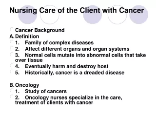
Indications for Cranial Surgery Brain Tumors • Steriotactical techniques used to perform biopsy and/or remove small tumors • Location and type determines if surgical removal possible • Tumors located in deep central areas of brain inoperable • Cranial surgery performed if tumor is removable
Brain Tumors (cont.) • Primary tumors - arise from tissues in the brain • Secondary tumors - result from metatastisis from malignant neoplasm elsewhere in body • Gliomas account for 65% of primary tumors (malignant) • Astrocytoma- most common glioma • Oligodendroglioma-often localized frontally • Glioblastoma multiforme highly malignant and invasive • Meningioma and Pituitary tumors • Benign • Tend to recur • Unless treated, all tumors cause death from increased tumor volume leading to increased ICP
Indications for Cranial Surgery Hydrocephalus • Overproduction, malabsorption, or accumulation of CSF. Shunting procedure performed to drain CSF.
Aneurysm Repair A clip is placed across the neck of the aneurysm which originates from the carotid artery
Preoperative nursing management • Preoperative teaching to patient and family • Explain preop labs, tests, procedures • Explain anesthesia, estimated length of procedure, how long in recovery and where will go after recovery (ICU) • Explain how pt. will look after surgery • Explain what to expect postoperatively re: dressings, catheter, ET tube, Foley, IV’s, IS, pain management
Preoperative nursing management (cont.) • Nearest relative may need to sign consent • Scalp prep - hair shaved (save hair) to reduce risk of infection and provide better exposure • Baseline neuro assessment • Family anxious re: potential physical and emotional deficits related to surgery - compassionate preoperative nursing care
Types of Cranial Surgery:Burr Hole-to remove blood/fluid or in preparation for a craniotomy
Craniotomy (cont.) After the dura has been stitched closed, the piece of bone is replaced and sutured into place. An ICP monitoring device may then be implanted.
Shunt Procedures While the patient is deep asleep and pain-free (using general anesthesia), a flap is cut into the scalp, and a small hole is drilled in the skull.
Shunt Procedures (cont.) A small catheter is passed into a ventricle of the brain. A pump is attached to the catheter to keep the fluid away from the brain. Another catheter is attached to the pump and tunneled under the skin, behind the ear, down the neck and chest, and into the peritoneal cavity (abdominal cavity). The CSF is absorbed in the peritoneal cavity.
Minimally Invasive Cranial Surgery A preoperative cerebral arteriogram (A) shows a basilar tip aneurysm. A postoperative arteriogram, after aneurysm clipping via a superolateral orbital craniotomy, confirms successful clipping (B). A patient with a healed superolateral orbital craniotomy incision line (C) (arrows).
Steriotaxis Advantages: • non-invasive • less risky than crani- otomy • decreased cost • decreased length of stay, recovery "stereotactic radio surgery”- removing tumors with radiation to a specific target, without radiating the entire brain
Nursing Management after Cranial Surgery • Primary Goal of Care - prevention of increased ICP • Ventriculostomy • Drains CSF • Allows for intraventricular drug administration • Measures pressure within vessels
Monitor ICP and CPP Pressure Waves A waves (plateau waves) - associated with ICP>20 - indicates exhausted intracranial spatial compensation - associated with increased cerebral volume and decreased cerebral blood flow, cerebral ischemia and brain damage
B waves - rhythmic oscillations approx. q min - associated with fluctuating breathing pattern C waves - associated with normal changes in systemic art. pressure B waves in raised ICP
Nursing management after cranial surgery (cont.) • Frequent assessment of neurological status (every 30 minutes, then hourly) for the first 24-48 hours • Frequent vital signs • Limit care activities that increase ICP • DO NOT cluster cares!
Elevate HOB 30 to 45 degrees for supratentorial surgery Keep patient flat or slightly elevated if incision in posterior fossa (infratentorial) Nursing management: Positioning
Nursing management after cranial surgery (cont.) • Assess for pain and provide pain relief measures-narcotics mask LOC • Check drains for placement, patency - strict sterile technique • Check dressing for drainage, CSF leak - strict sterile technique • Suction—limit to < 15 seconds; preoxygenate • Turn q 2 hrs (slow, gentle movements) • ROM exercises
Nursing management after cranial surgery (cont.) • Assess effect of ill family member on family • Teach family to provide care to ill family member • Facilitate family communication and planning • Provide accurate information to family regarding patient’s condition • Initiate referrals as needed, i.e. speech therapy, physical therapy
Postoperative Medications • Anticonvulsants • Corticosteroids • Histamine blockers • Analgesics • Antibiotics
Postoperative Complications • Increased intracranial pressure (ICP) • Hematomas Subdural hematoma Epidural hematoma Subarachnoid hemorrhage
Postoperative complications (cont.) • Hypovolemic shock • Hydrocephalus • Respiratory Complications Atelectasis Hypoxia Pneumonia Neurogenic pulmonary edema
Postoperative Complications (cont.) • Infection • Meningitis • Fluid and electrolyte imbalances • Dehydration • Hyponatremia • Hypernatremia
Postoperative Complications (cont.) • Seizures • Cerebrospinal fluid (CSF) leak • Cerebral edema
Summary • Neuro care complex • Encompasses science and art of nursing • Requires technical expertise • Requires collaboration, communication, compassion