Download
1 / 23
240 likes | 277 Views
Learn about the methods for diagnosing fungal diseases, including sample collection, examination, and culture techniques. Follow laboratory safety rules to prevent contamination and ensure proper handling of infectious materials.

E N D
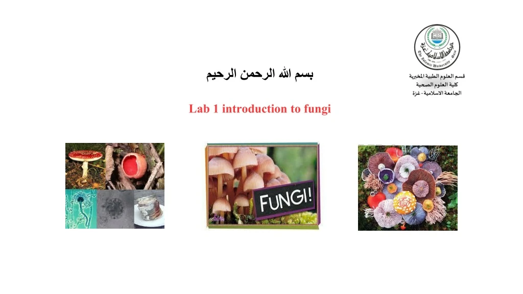
بسم الله الرحمن الرحيم Lab 1 introduction to fungi
Introduction • Medicinal fungi: it is the fungi that cause diseases to humans and animals resulting from the toxic substances they produce, this causes a major disruption to the cellular system, causing disruption to its functioning, leading to a disease . • There are many methods for collection of samples and procedures for diagnosis to determine the causative agent of the disease.
One of the fastest ways to identify the causative agent is direct examination of the sample. • In order to confirm the preliminary diagnosis, sample is cultured in suitable medium. • Samples are taken as swabs or tissue sections and diagnosed by wet mount, KOH smear, staining ,etc.
Samples 1) Nail. 2) Skin. 3) Hair. 4) CSF. 5) Urine. 6) Vaginal secretions. 7) Respiratory specimen. 8) Bone marrow. 9) Blood. 10) Tissue biopsy.
Fungus culture • The pathogenic fungi are cultured in petri dishes containing media as Sabroud Agar which is one of the most important media used for in the diagnosis. • Then dishes will be incubated at 25 ° C and the fungal cultures will be examined by: 1) Macroscopic: Developing colonies are monitored during the first week of incubation. At the end of the third week, notes are recorded and include:
A. Growth. B. General appearance: The colonies are flat, rounded, Regular or irregular. C. The structure of the colonies is as soft as yeast, smooth, grainy, Velvet, cotton, Hey.
E. Pigmentation: Fungi may be colorless or brightly colored. • Color may be on fungus itself, on its sporulating apparatus, on the agar, or on the bottom of the colony (reverse pigmentation). • Pigment color is due to the color of the sporulating apparatus. • The pigment can be diffused into the agar. • It is important to note the top pigment (obverse) and the discoloration of the agar medium (reverse).
2 .Microscopic: • A small part of the colony is taken by the loop with a small drop of lactophenol blue or small drop of normal saline on a slide. • Next, place the cover slip and examine below the microscope for the detection of hypha mycelium and spores.
Yeast: It is a unicellular organism that reproduces by budding or by binary fission. • Hypha: elongation of apical cell produces a tubular, thread like structure called hypha. Hypha may be septate or non septate. • Mycelium: tangled mass of hypha is called mycelium. • Fungi producing mycelia are called molds or filamentous fungi.
Rules General lab. Safety • In the microbiology laboratory infectious materials are processed as many of the microorganisms used in this course may be pathogenic for humans . • Also the materials found, such as glass equipment, biological agents and chemicals can pose safety hazards to you if you do not follow laboratory protocols. • As a result, certain rules are necessary to prevent the spread of infectious agents, the possibility of infecting yourself or other people and to prevent contamination of specimen with environmental microorganisms.
The following laboratory safety rules must be followed : • All health-care workers should routinely use appropriate barrier precautions to prevent skin and mucous-membrane exposure when contact with blood or other body fluids of any patient • Safety in a microbiology laboratory starts with protecting yourself : • cover long hair or keep it tied up and out of way. • Always wearing closed shoes. • Wearing personal protective equipment (coat, gloves, eye protection; glasses may be preferred to contact lenses).
Never applying cosmetics, or placing objects (fingers, pencils) in the mouth or touching the face. • Never eating, drinking or chewing gum in the laboratory. • For working : • Washing hands prior and after lab working with water and soap. • Keep the lab bench free of unnecessary materials. • Disinfecting lab benches prior to and at the conclusion of each lab session.
Dispose all contaminated materials in autoclave bags and proper disposal of other different types of waste. • The coats should be kept separately from other things in your bag. • Reporting all injuries, accidents, spills and broken glassware to the instructor and receiving instructions for cleanup. • Always using appropriate pipetting devices and understanding that mouth pipetting is forbidden. • Using universal precautions posted in the lab and see inside front cover of this laboratory manual.
Safety for dealing with specimen: • Blood and other body fluids from all patients should be considered infective. • All health-care workers should take precautions to prevent injuries caused by needles, scalpels, and other sharp instruments or devices during procedures. • To prevent needle stick injuries, needles should not be recapped.
Avoid contaminating the outside of the container. • All specimens should be put in a well-constructed container with a secure lid to prevent leaking during transport. • After they are used, disposable syringes and needles, scalpel blades, and other sharp items should be placed in puncture-resistant containers for disposal.
Decontamination of small & moderate spills • Notify other workers in the area of the spill and control traffic through area. • Wear shoe covers and safety goggles, if spill is on floor may have splashed beyond immediate area of spill. • Put on gloves and cover spill area with paper towels. • Pour disinfectant over towels from edges of spill to center, be carefully not to splatter.
Decontaminate all objects in spill area. • Allow 30 minutes of contact time. • Pick up any sharps, including broken glass with forceps and place in sharps container. • Use squeegee and dustpan to recover any shards of broken glass in contaminated liquid. Decontaminate squeegee and dustpan. • Wipe area with disinfectant and clean paper towels and put in biohazard bag.
Mop if spill is in floor. • Remove gloves and shoes cover before removing area of the spill, put on biohazard bag, Wash hands. • Decontamination of large spills : • Evacuate room, close doors, prevent others from entering and wait 30 minutes for aerosols to settle. • Follow previous procedure for small and moderate spills.
Lab requirements • Lab coat • Matches or lighter • Soap • Waterproof permanent marker • Small or medium size towel • Sealable plastic bag • Rubber band
Instruments in mycology lab • incubators • Autoclave
Benzene Burner Safety Cabinet


