Download
1 / 29

E N D
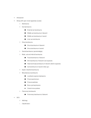
Introduction • Diving with open circuit apparatus (scuba) • Barotrauma ○ Ear barotrauma ○ External ear barotrauma Middle ear barotrauma of descent Middle ear barotrauma of ascent Inner ear barotrauma Sinus barotrauma ○ Sinus barotrauma of descent Sinus barotrauma of ascent Dental barotrauma (aerodontalgia) ○ Mask, suit and helmet barotrauma ○ Facial barotrauma of descent Skin barotrauma of descent (suit squeeze) Head and body barotrauma of descent (diver's squeeze) Suit barotrauma of ascent (‘blow up’) Gastro intestinal barotrauma ○ Miscellaneous barotrauma ○ Localised surgical emphysema Pneumoperitoneum Pneumocephalus Bone cyst barotrauma Cranial nerve palsies Pulmonary barotrauma ○ Pulmonary barotrauma of descent DCS • Aetiology ○ Classification ○
Onset ○ Generalised ○ Muskuloskeletal ○ Neurological ○ Cutaneous ○ Gastrointestinal ○ Cardiorespiratory ○ Treatment ○ • Dysbaric osteonecrosis Type A lesions ○ Type B lesions ○ Salt water aspiration syndrome • Clinical ○ Headaches after diving differential diagnosis • Recommended reading • Introduction The illnesses encountered in diving medicine are related to the environment and/or the diving equipment. Thus it is conventional to subdivide the divingrelated illnesses as to whether they occur during free diving, with open circuit diving, or with closed circuit diving equipment. The freediving hazards are due mostly to the aquatic environment, and therefore are common to all types of divers. Swimming and breathhold diving accidents include: (i) the drowning syndromes (ii) the effects of cold and immersion (iii) marine animal injuries (iv) hyperventilation then breathholding hypoxia (v) descent barotrauma. Apart from descent barotrauma, these disorders are well described in general medical texts and in the more comprehensive diving medical texts.
The hazards associated with open circuit equipment are commonly encountered, as they include diving The hazards associated with open circuit equipment are commonly encountered, as they include diving with scuba. The diseases are especially those of the extended environmental parameters, to which the equipment now allows the diver to explore. Thus the illnesses of barotrauma, decompression sickness, bone necrosis and gas toxicities develop. This chapter deals essentially with the dysbaric diseases (due to abnormal environmental pressures) as well as some of the common symptoms with which divers present (hearing loss, vertigo, headaches). The hazards with rebreathing equipment tend to be restricted to professional or technical divers, and their medical backup. They are more related to the extremes of gas pressures (especially oxygen, carbon dioxide, nitrogen and helium), and they develop because of the complexity of maintaining breathing gas pressures within an acceptable range for humans. Despite the above, many of the accidents experienced by divers are initiated because of the medical illnesses and personal characteristics of the diver (inadequate physical fitness, psychological disturbances, inadequate training etc.) and improper or unsafe diving techniques and practices. These are well covered in the conventional diving medical texts, and do not usually present as the problem in emergency medical situations. They are the provoking factors on which the medical disease will develop. Diving with open circuit apparatus (scuba) This equipment allows the inhalation of gases from a high pressure source, and the liberation of exhaled gas into the water. There is no rebreathing of exhaled gases involved. Open circuit diving may involve the use of: i. Self contained underwater breathing apparatus (scuba). The breathing gas is compressed air and is carried in tanks on the diver's back. ii. Surface supply breathing apparatus (SSBA). Air is carried to the diver via a hose from either storage cylinders or compressors (‘hookah’) on the surface. iii. Standard diving equipment (‘hard hat’, etc.). Gas mixtures (oxygen/nitrogen, oxygen/helium) may replace air for specific diving operations The availability of nonexpensive equipment has increased the popularity of this type of diving. Scuba and SSBA have largely replaced the old ‘hard hat’ or standard diving previously used in shell collecting and salvage. The medical conditions which can occur when diving with compressed gases include: Barotrauma ears, sinus, lung, dental, others. i. ii. Decompression sickness. Dysbaric osteonecrosis iii.
iv. Salt water aspiration. v. Gas toxicities. vi. Contamination of gas supply. vii. Nitrogen narcosis. viii. Syncope of ascent. Only the first four of these are likely to present to the emergency room clinician, and will be dealt with here. Common diving medical presentations have been included, with a check list to aid in differential diagnosis: i. Disorientation and Vertigo in diving. ii. Hearing loss iii. Headaches Physics review 1 To understand the physiological changes which occur, some knowledge of physics is needed. Pascal's Principle states that when pressure is applied to the surface of a fluid it is distributed equally and undiminished in all directions. Since the pressure on sea water is atmospheric pressure — that is, 100 kPa , (1 Bar, 1 atmosphere absolute, or 1 ATA)— then at a depth of 10 metre the pressure will be 1 ATA plus the pressure equal to 10 m of sea water. In sea water, the pressure will increase by 100 kPa (1 ATA) for every 10 m depth. The pressure at 10 m will then be as follows; 1 ATA + 1 ATA = 2 ATA. Surface pressure = 1 ATA At 10 m = 2 ATA At 20 m = 3 ATA At 30 m = 4 ATA Boyle's Law states that at a given temperature, the volume of a given mass of gas will vary inversely with the pressure applied. PV = K (where K is a constant). This means that 1 litre of gas at the surface (where the pressure equals 1 ATA) will be reduced to 0.5 I at 10 m (2 ATA), or 1/3 I. at 20 m depth (3 ATA), 1/4 I. at 30 m depth (4 ATA) etc. It can be seen that the volume change is proportionately greatest near the surface, and so it is in this
zone that the effects of Boyle's Law are most noticeable. zone that the effects of Boyle's Law are most noticeable. Barotrauma Barotrauma is the tissue damage resulting from the expansion or contraction of enclosed gas spaces, and is a direct effect of the gas volume changes causing tissue distortion. It is probably the most common occupational disease of divers. There are two types of barotrauma descent and ascent. Both are caused by the effects of Boyle's Law. The volume change is proportionally greatest near the surface, and so it is here that barotrauma is most noticeable. Barotrauma of descent is that damage which occurs during descent in water, i.e. as a result of increasing pressures of the surrounding environment. Pressure imbalance is due to an inability to compensate for the reducing volumes within the body cavities as the depth increases. Because some cavities are surrounded by bone, no collapse can occur, and the space must be taken up by engorgement of the mucous membrane, oedema and haemorrhage. This, together with the compressed gas, assists in ‘equalising’ the pressure balance. It is commonly called a ‘squeeze’. Barotrauma of ascent is the result of the distension of tissues around the expanding gas. This occurs when environmental pressures are reduced during ascent in water. Divers use the misnomer ‘reverse squeeze’ to describe it. The classification of barotrauma is as follows : EAR BAROTRAUMA External ear barotrauma of descent Middle ear barotrauma of descent Middle ear barotrauma of ascent Inner ear barotrauma SINUS BAROTRAUMA Sinus barotrauma of descent Sinus barotrauma of ascent DENTAL BAROTRAUMA MASK, SUIT AND HELMET BAROTRAUMA Facial barotrauma of descent Skin barotrauma of descent Head and body barotrauma of descent Suit barotrauma of ascent GASTRO INTESTINAL BAROTRAUMA
OTHER BAROTRAUMA AND SEQUELAE Localised surgical emphysema Bone cyst Pneumoperitoneum Pneumocephalus Cranial nerve palsies Ear barotrauma This is the commonest reason for divers to present to clinicians. Barotrauma is subdivided according to the anatomical sites. They may occur separately or in combination in the external ear, the middle ear, or the inner ear. External ear barotrauma (external ear squeeze, reversed ear). If the external meatus is blocked, water entry is prevented. Then contraction of the contained gas during descent is compensated for by tissue collapse, outward bulging of the tympanic membrane, congestion and haemorrhage. These results are observed in as little as 2 metres of water. The common causes of blockage of the external auditory canal include; cerumen, exostoses, foreign bodies such as mechanical ear plugs, tight fitting hoods and mask straps. Clinical symptoms are usually mild. Following ascent there may be an ache in the affected ear and/or a bloody discharge. Examination of the external auditory canal may reveal petechial haemorrhages and bloodfilled cutaneous blebs which may extend onto the tympanic membrane. Treatment for this condition includes maintaining a dry canal, removal of any occlusion and prohibition of diving until epithelial surfaces appear normal. Secondary infection may result in a recurrence of the pain, and require antibiotics. This condition is easily prevented by ensuring patency of external auditory canals and avoidance of ear plugs or hoods which do not have apertures over the ear to permit water entry. Middle ear barotrauma of descent (middle ear squeeze) This is the most common medical disorder experienced by divers, and it follows the failure to equalise middle ear and environmental pressures via the Eustachian tubes (‘equalising the ears’), during descent. Any condition which blocks the Eustachian tube, predisposes to middle ear barotrauma. Failure to voluntarily autoinflate the middle ears, usually by the Valsalva manoeuvre during descent, has the same effect. If the diver continues the descent without ‘equalising’, mucosal congestion, oedema and haemorrhage within the middle ear cavity are associated with inward bulging of the tympanic membrane. This tends to compensate for the contraction of air within the otherwise rigid cavity. The tympanic membrane will become haemorrhagic ( the ‘traumatic tympanum’ of older texts). Eventually it may rupture, although this is not common.
Blockage of the Eustachian tubes may be due to mucosal congestion as a manifestation of upper Blockage of the Eustachian tubes may be due to mucosal congestion as a manifestation of upper respiratory tract infections, smoking, allergies, otitis media, mechanical obstructions such as mucosal polyps, or individual variations in size, shape and patency. Symptoms consist initially of discomfort followed by increasing pain in the ear if descent continues. This may be sufficiently severe to prevent further descent. Occasionally a diver may have little or no symptomatology. Occasionally, there is a sensation of vertigo during the descent, but it is not as common as in middle ear barotrauma of ascent or inner ear barotrauma (see later), both of which can follow or be due to middle ear barotrauma of descent. Eventually rupture of the drum may occur, usually after a descent of 1.510 metres from the surface. This causes instant equalisation of pressures by allowing water entry into the middle ear cavity. If this occurs, pain is suddenly relieved; however, nausea and vertigo may follow the caloric stimulation by the cold water. Following a dive which has resulted in descent barotrauma, there may be a mild residual pain in the affected ear. Blood or blood stained fluid may be expelled from the middle ear during ascent, and be swallowed or produce epistaxis on the affected side. A full or blocked sensation may be experienced in the ear. This is sometimes associated with a mild conductive deafness especially involving low frequencies. It is usually temporary. Fluid may be felt within the middle ear for a week or so, before resolution. Middle ear barotrauma is classified into six grades based on the otoscopic appearance of the tympanic membrane. The grades are: MIDDLE EAR BAROTRAUMA OF DESCENT - GRADING. GRADE 0 - Symptoms without signs; GRADE I - Injection of the tympanic membrane, especially along the handle of the malleus; GRADE II - Injection plus slight haemorrhage within the substance of the tympanic membrane; GRADE III - Gross haemorrhage within the substance of the tympanic membrane; GRADE IV - Free blood in the middle ear as evidenced by blueness and bulging; GRADE V - Perforation of the tympanic membrane. Damage involves the whole of the middle ear space and not the tympanic membrane alone. The clinical management consists of: • prohibition of all pressure changes such as diving and autoinflation techniques until resolution;
• occasionally (very rarely) systemic or local decongestants; • antibiotics only where there is evidence of a pre-existing or developing infection, gross haemorrhage or perforation. Serial audiometric examination should be undertaken to exclude hearing loss, and to assist in further action if such loss is present. Diving can be resumed when resolution is complete, and voluntary autoinflation of the middle ear cleft has been demonstrated. If there is no perforation (grades 0-4), recovery may take up to two weeks. With perforation (grade 5) it may take one to two months, if uncomplicated and managed conservatively . It is important to clearly identify the contributing factors to the disease in each case, or it is likely to recur. Middle ear barotrauma of ascent This refers to the effects from distension by enclosed gases within the middle ear, expanding with ascent. Because it may prevent ascent, it is usually considered more serious than middle ear barotrauma of descent - which allows an unhindered return to safety. Gas which has entered the middle ear by autoinflation at depth is at the surrounding environmental pressure, and on ascent it obeys Boyle's Law. If the Eustachian tube restricts its release, the expansion of gas can cause clinical manifestations with sensations of pressure or pain in the affected ear, vertigo due to increased middle ear pressure difference (alternobaric vertigo), or tinnitus. The vertigo tends to develop when the middle ear's pressure differs by 60 cm H2O. Relief of the overpressure in the affected middle ear may be heard, with air felt hissing out the Eustachian tube. Hearing loss in the affected ear, if present, may be either conductive or sensorineural and may follow damage to the tympanic membrane or the middle ear structures. Inner ear barotrauma is a possible complication. Seventh nerve palsy also is a complication (see later). Middle ear barotrauma of ascent usually follows recent, but sometimes mild, middle ear barotrauma of descent or the use of nasal decongestants. In each case the common factor is probably a congestion and therefore blockage of the Eustachian tube. Otoscopic examination often reveals evidence of tympanic membrane injection or haemorrhage. Treatment is as for middle ear barotrauma of descent. Prevention is achieved by avoiding nasal decongestants and by training the diver in correct middle ear equalisation techniques. Unless the descent barotrauma is prevented the ascent barotrauma is likely to recur. Inner ear barotrauma There is always the possibility of a sensorineural hearing loss in divers who have experienced ear barotrauma of any type, or who have had difficulty in equalising middle ear pressures by autoinflation or
who subsequently apply force to achieve this. In these cases, the hearing loss may immediately follow the incident, or may develop over the next few days. An inner ear (labyrinthine) window fistula is one pathological entity of inner ear barotrauma. Others include cochlear and vestibular haemorrhages, internal inner ear membrane ruptures, the entry of air into the cochlea, etc. It has been reported from dives as shallow as 7 feet (2 metres) and has been observed in a surfer who merely dived under a wave. Symptoms associated with inner ear barotrauma may include; • a sensation of blockage in the affected ear • tinnitus of variable duration • high frequency hearing loss • vestibular disturbances such as nausea, vomiting, vertigo, disorientation and ataxia • clinical features of an associated middle ear barotrauma . Any combination of middle ear barotrauma symptoms, vertigo, tinnitus and hearing loss should be immediately and fully investigated by serial measurements of clinical function, daily audiometry up to 8000 Hz and positional electronystagmography. Caloric testing is indicated only if the tympanic membrane is intact or if the technique guards against pressure or fluid transmission into the middle ear. Other investigations that may be of value include temporal bone polytomography, CT scans and other imaging techniques. Until now they have not been particularly helpful in diagnosis or treatment, but this should change with more experience. Cochlear injury is permanent in over half the cases, whereas vestibular injury is usually temporary. Treatment. Once damage has been confirmed, treatment should be initiated promptly. This includes: i. Avoid straining, such as performing Valsalva manoeuvres, sneezing, nose blowing, straining with defaecation, sexual activity, coughing, lifting weights or physical exertion. Any increase in cerebrospinal fluid pressure can be transmitted to the inner ear. ii. Immediate bed rest with the head elevated and careful monitoring of otological changes; this is given irrespective of which of the other treatment procedures are followed. iii. Bed rest should continue until all improvement has ceased and for up to a week thereafter, to allow the inner ear membranes to heal and the haemorrhages to resolve. Loud noises should be avoided. iv. If there is no improvement within 24 - 48 hours in cases of severe hearing loss, or if there is further deterioration in hearing, reconstructive microaural surgery must then be considered. v. Prohibition of diving and flying. This is absolute for the first few weeks following a labyrinthine window fistula. If medical evacuation by air is required, an aircraft with the cabin pressurised to ground level is necessary. Hyperbaric oxygen therapy has been used in some cases, but requires further confirmation before it can
be generally recommended. Sinus barotrauma If a sinus ostium is blocked during descent, mucosal congestion and haemorrhage compensate for the contraction of the air within the sinus cavity. During ascent, expansion of the enclosed air expels blood and mucus from the sinus ostium. Ostia blockage may be the result of sinusitis with mucosal hypertrophy and congestion, rhinitis, redundant mucosal folds in the nose, nasal polyps etc. Allergies and smoking may underly the pathology. Sinus barotrauma of descent (sinus squeeze) Symptoms include pain over the sinus during descent. It may be preceded by a sensation of tightness or pressure. The pain usually subsides with ascent but may continue as a persistent dull ache for several hours. On ascent blood or mucus may appear in the nose or pharynx. The pain is usually over the frontal sinus, less frequently it is retro-orbital, and maxillary pain is not common but may be referred to a number of upper teeth. Although they may feel hypersensitive, abnormal or loose, they are not painful on movement. Numbness over the maxillary division of the trigeminal nerve is possible. The superficial ethmoidal sinuses near the root of the nose occasionally rupture and cause a small haematoma or discolouration of the skin, between the eyes. Discomfort persisting after the dive may be due to fluid within the sinus (remaining from the dive), infection (usually starts a few hours post-dive) or the development of chronic sinusitis or mucoceles. Sinus x-ray examination, CT or MRI scan may disclose thickened mucosa, opacity or fluid levels. The opacities produced by the barotrauma may be serous or mucous cysts. The maxillary and frontal sinuses are commonly involved. The ethmoid and sphenoidal sinuses may also be affected. The new imaging techniques can clearly demonstrate these. Treatment consists of temporary cessation of all diving and flying, with correction of any predisposing factors. Patients with a sinus or upper respiratory tract infection may require antibiotics and decongestants. Surgical drainage is rarely indicated. Even the mucoceles and chronic sinus pathology usually resolve without intervention, if diving is suspended. Sinus barotrauma of ascent This may follow the occlusion of sinus openings by mucosal folds or sinus polyps, preventing escape of expanding gases. The ostium or its mucosa will then blow out into the nasal cavity, with or without pain, and haemorrhage commonly follows. This disease is aggravated by rapid ascent. If the expanding air cannot escape through the sinuses it may fracture the walls and track along the soft
tissues. Rupture of air cells may cause sudden pain of a severe degree, often affecting the ethmoidal or mastoid sinuses, on ascent. Occasionally the air may rupture into the cranial cavity and cause a pneumocephalus. Dental barotrauma (aerodontalgia) The tooth may cave in (implode) on descent, or explode on ascent. Gas spaces may exist in carious teeth, at the roots of infected teeth or within fillings which have undergone secondary erosion. During descent, the gas space contracts, filling with soft tissue or blood. Pain may prevent further descent. Gas expansion on ascent may be restricted by blood in these spaces, resulting in pain which may continue post-dive. Pressure applied to individual teeth may cause pain and identify the affected tooth. Treatment consists of analgesia and dental repair. The differential diagnosis of sporadic or constant pain in the upper bicuspids or the first and second molars, but not localised in one tooth, must include referred pain from the maxillary sinus or the maxillary nerve. This may also present as a burning sensation along the mucobuccal fold. Mask, suit and helmet barotrauma Facial barotrauma of descent (mask squeeze) A face mask creates an additional gas space external to, but in contact with, the face. Unless pressure is equalised by exhaling gas through the nose, facial tissues will be forced into this space, during descent. Clinical features include puffy oedematous facial tissues especially under the eyelids, purpuric haemorrhages, conjunctival haemorrhages, and later, generalised bruising of the skin underlying the mask. This condition is rarely serious, and prevention involves exhaling into the face mask during descent. Diving should be avoided until all tissue damage is healed. Rare cases may involve the deeper orbital tissues and/or retina. Skin barotrauma of descent (suit squeeze) This condition is encountered mainly with dry suits or poorly fitting wet suits. During descent the air spaces are reduced in volume and trapped in folds in the suit. The skin tends to be sucked into these folds, leaving linear weal marks or bruises. The condition is usually painless and clears within a few days. Head and body barotrauma of descent (diver's squeeze) A rigid helmet, as used in standard and ‘hard hat’ diving, may cause this trauma. If extra gas is not added during descent to compensate for the effects of Boyle's Law, or if pressure is lost for any reason, the suit and occupant may be forced into the helmet, causing fractured clavicles, bizarre injuries or
death. Suit barotrauma of ascent (‘blow up’) During ascent with a gas filled diving suit (a ‘dry suit’), the expanding gas must be able to escape. If it does not, then the whole suit will expand like a balloon and cause a rapid and uncontrolled ascent to the surface. This may result in barotrauma of ascent, decompression sickness, imprisonment of the diver and physical trauma. Gastro intestinal barotrauma Gas expansion occurs within the intestines on ascent, and may result in eructation, vomiting, flatus, abdominal discomfort and colicky pains. It is rarely severe, but has been known to cause syncopal and shock-like states. A small group of divers have experienced more serious symptoms, with gastric rupture on ascent. Miscellaneous barotrauma Localised surgical emphysema This may result from the entry of gas into any area where the integument, skin or mucosa is broken and in contact with a gas space. Although the classical site involves the supraclavicular areas in association with tracking mediastinal emphysema from pulmonary barotrauma, other sites are possible. Orbital surgical emphysema, severe enough to completely occlude the palpebral fissure, may result from diving with facial skin, intra-nasal or sinus injuries. The most common cause is a fracture of the naso- ethmoid bones. The lamina papyracea, which separates the nasal cavity and the orbit, is of egg shell thickness. When these bones are fractured, any increase in pressure in the nasal cavity or ethmoidal sinus from ascent or Valsalva manoeuvre, may force air into the orbit. Surgical emphysema over the mandibular area is common with buccal and dental lesions. The surgical emphysema, with its associated physical sign of crepitus and its radiological verification, tends to occur in loose subcutaneous tissue. Treatment is by administration of 100% oxygen by a non-pressurised technique, and complete resolution will occur within hours. Recompression is rarely indicated, but diving should be avoided until this resolution is complete and the damaged integument has completely healed. Pneumoperitoneum This has been observed following pulmonary barotrauma, gas dissecting along the mediastinum to the retroperitoneal area, into the peritoneum, and under the diaphragm. It is also possible that previous injury to the lung or diaphragm, producing adhesions, could permit the direct passage of air from the lung to the subdiaphragmatic area.
Another possible cause of pneumoperitoneum is, as described above, from a rupture of a gastrointestinal viscus - from barotrauma of ascent. The condition may be detected by chest x-ray or positional abdominal x-ray (gas under the diaphragm). Treatment is by administration of 100% oxygen by a non-pressurised technique. Usually complete resolution will then occur within hours. Management of the cause (pulmonary or gastro-intestinal) is required and surgical management of a ruptured gastrointestinal viscus may be needed. Pneumocephalus Occasionally the cranial gas spaces (mastoid, para-nasal sinuses) are affected by an ascent barotrauma, when the expanding gas ruptures into the cranial cavity. This may follow descent barotrauma, when haemorrhage occupies the gas space and its orifice is blocked. The clinical presentation may have all the features of a catastrophic intracerebral event. Excruciating headache immediately on ascent is probable, although the effects of a space occupying lesion may supervene. Neurological signs may follow brain injury or cranial nerve lesions. It is likely that the condition could be aggravated by excessive Valsalva manoeuvres (‘equalising the ears’) or ascent to altitude (air travel). Diagnosis can be verified by positional skull x-ray, or CT scan. Treatment includes: bed rest, sitting upright; avoidance of Valsalva, sneezing, nose blowing or other manoeuvres that increase nasopharyngeal pressures; 100 % oxygen inhalation for many hours and follow up x-rays to show a reduction of the air volume. If untreated, the disorder may last a week or so and subsequent infection is possible. Recompression or craniotomy could be considered in dire circumstances. Bone cyst barotrauma Occasionally pain may develop from an intra-osseous bone cyst, probably with haemorrhage into the area, during descent or ascent, and may last for hours after the dive. The pelvic bones are most often involved, in the ilium and near the sacroiliac joints. An x-ray or CT scan may demonstrate the lesions. Cranial nerve palsies Cases occasionally present with cranial nerve lesions attributed to neurapraxis, due to the implosive tissue damaging effects during descent, the distension in enclosed gas spaces during ascent, or both. These presentations are usually associated with barotrauma symptoms and signs, as described earlier. The seventh or facial cranial nerve may be affected, causing ‘facial baroparesis’, because of its passage through the middle ear space. Recorded in both aviators and divers, it is more frequent following ascent, presents as a unilateral facial weakness similar to Bell's palsy, and tends to recur in the same patient. A possible reason for an individual's susceptibility to this disorder is perhaps found in the anatomy of the facial canal. This opens into the middle ear in some people and shares its pathology. The fifth or trigeminal nerve may be likewise influenced by gas pressure changes in the maxillary antrum
and sinus barotrauma (see above). The most common presentation is with involvement of the maxillary division, especially the infraorbital nerve, which traverses the sinus. Hypoaesthesia can be demonstrated for a variable time after the sinus barotrauma incident. It may involve the cheek, side of nose, lower eyelid, upper lip, maxillary teeth and gums. It may also be a cause of pain from sinus barotrauma referred to the upper teeth on the same side. Pulmonary barotrauma Pulmonary Barotrauma (PBT) of ascent is the most serious of the barotraumata, and causes concern in all types of compressed air diving. It is the clinical manifestation of Boyle's Law as it affects the lungs and is the result of over distension and rupture of the lungs by expanding gases during ascent. It is also called ‘burst lung’ or pulmonary overinflation. It is second only to drowning as a cause of death amongst young recreational scuba divers. Controversy surrounds the pathophysiology of PBT, but its cause, diagnosis and treatment are well established. Aetiology. PBT may involve much of the lung, such as when the expanding lung gases are not exhaled during ascent. Alternately it may involve only small areas following obstructed air flow or altered compliance in some airways. With a breathhold ascent, the pressure change necessary to cause PBT is approximately 70 mm Hg near the surface, i.e. a force which could cause an increase in lung volume of about 10% i.e. with an ascent from a depth of about 1 metre, to the surface. Predisposing pathology causing local ruptures include previous spontaneous pneumothorax, asthma, sarcoidosis, cysts, tumours, pleural adhesions, intra pulmonary fibrosis, infection and inflammation, etc. These disorders may result in local compliance changes or airway obstructions. There are four manifestations of PBT of ascent which may occur singly or in combination: • Pulmonary tissue damage; • Mediastinal emphysema; • Pneumothorax; • Air embolism. Clinical features. Pulmonary tissue damage. Dyspnoea, cough and haemoptysis are symptoms of the lung damage, and widespread alveolar rupture may cause death from respiratory damage. Mediastinal Emphysema. Symptoms may appear rapidly in severe cases, or may be delayed several hours in lesser cases. They may include a voice change into a hoarseness or a brassy monotone, a feeling of fullness in the throat, dyspnoea, dysphagia, retrosternal discomfort, syncope, shock or unconsciousness. The voice changes are attributed to ‘submucosal emphysema’ of the upper airways and/or recurrent laryngeal nerve damage.
Clinical signs include: subcutaneous emphysema of neck and upper chest wall, i.e. crepitus under the skin (described as the sensation of egg-shell crackling, by divers), decreased areas of cardiac dullness to percussion, faint heart sounds, left recurrent laryngeal nerve paresis, and cardiovascular effects of cyanosis, tachycardia and hypotension. Precordial emphysema may be palpable and give the pneumoprecordium or Hamman's sign - crepitus related to heart sounds. An extension of the mediastinal gas into the tissues between the pleura and the pericardium, rather than gas in the pericardial sac, has produced cardiac tamponade with its clinical signs. There may be radiological evidence of an enlarged mediastinum with air tracking along the cardiac border or in the neck. Pneumothorax. If the visceral pleura ruptures, air enters the pleural cavity and expands during further ascent. It may be accompanied by haemorrhage, forming a haemopneumothorax. The pneumothorax may be unilateral or bilateral. Symptoms usually have a rapid onset and include sudden retrosternal or unilateral, sometimes pleuritic, pain with dyspnoea and an increased respiratory rate. Clinical signs may be absent, or may include: diminished chest wall movements, diminished breath sounds and hyper resonance on the affected side; movement of trachea and apex beat to the unaffected side with a tension pneumothorax; signs of shock; x-ray evidence of pneumothorax; arterial gas and lung volume changes. A pneumothorax under pressure becomes a tension pneumothorax during ascent. Tension pneumothorax may also develop from coughing or exposure to altitude or aviation. Rarely a pneumoperitoneum may accompany the pneumothorax. Air (gas) embolism. This is a dangerous condition and is the result of gas passing from the ruptured lung into the pulmonary veins and thence into the systemic circulation, where it can cause vascular damage or obstruction, hypoxia and infarctions. Serious effects may result from blockage of cerebral (cerebral arterial gas embolism or CAGE) or coronary vessels by bubbles 25 microns - 2 mm in diameter, or by otherwise interrupting blood flow (vascular endothelial and perfusion injury). Death may follow coronary or cerebrovascular embolism. Other tissues may be affected. The manifestations are usually acute and may include: ○ Loss of consciousness; other neurological abnormalities such as confusion, aphasia, visual disturbances, paraesthesiae or sensory abnormalities, vertigo, convulsions, varying degrees of paresis; gas bubbles in retinal vessels; abnormal electroencephalograms and brain scans, etc. ○ Cardiac-type chest pain and/or abnormal electrocardiograms (ischaemic myocardium, dysrhythmias or cardiac failure) ○ Skin marbling; a sharply defined area of pallor on the tongue (Liebermeister's sign ) is
rare. Of divers who experience symptoms of CAGE, many will show a partial, or even a complete, recovery within minutes or a few hours of the incident. This recovery presumably reflects a movement of the embolus through the cerebral vasculature. Even those who become comatose may improve to a variable degree after the initial episode. Unfortunately the recovery is unreliable. It may not occur, or it may not be sustained. Recurrence of symptoms has an ominous prognostic significance. Treatment. Once PBT has resulted in the distribution of gas within body tissues, it may be aggravated by other factors. Further ascent in a chamber or underwater, or ascent to altitude during air transport, will expand the enclosed gas and cause deterioration in the clinical state of the patient. Physical exertion, increased respiratory activity, breathing against a resistance, coughing, Valsalva manoeuvre, etc. may also result in further pulmonary damage, or in more extraneous gas passing through the lung tissues or into the pulmonary vessels. If the diver has exposed himself to depths and times resulting in tissue loading by inert gas, this gas will diffuse into the abnormal gas spaces. A situation develops which has aspects of both PBT and decompression sickness. If the anaesthetic nitrous oxide is used, this rapidly diffuses into tissues, causing expansion of bubbles. Pulmonary tissue damage. The treatment is similar to that of near-drowning or the acute respiratory distress syndrome. Mediastinal emphysema. The need for therapy may not be urgent. Exclusion of air embolism or pneumothorax is necessary and if in doubt, treatment for these should take precedence. Management of mediastinal emphysema varies according to the clinical severity. If the patient is asymptomatic, only observation and rest may be necessary. With mild symptoms, 100% oxygen administered by mask without positive pressure will increase the gradient for removal of nitrogen from the emphysematous areas. This may take 4-6 hours. If symptoms are severe, therapeutic recompression using oxygen is necessary. Pneumothorax. Treatment depends on the clinical severity and the depth at which it is diagnosed. Any associated air embolism must be excluded. Mild cases require only administration of 100% oxygen, without positive pressure, bed rest and analgesics. A pneumothorax may also respond rapidly to high oxygen pressures at depth - such as when it occurs in a compression chamber or when a patient is recompressed for other reasons, such as CAGE. Serious cases, often with more than 20% lung collapse, may need to have the gas removed rapidly, by needle aspiration and/or intercostal cannulation and underwater drainage or a Heimlich valve, with or without low pressure suction. This may be needed while the patient is undergoing recompression therapy. Air Embolism. Treatment of air embolism is urgent, must be instituted immediately, and usually takes precedence over other manifestations of PBT. Immediate recompression is necessary, and a
recompression chamber should always be available. To reduce the likelihood of further CAGE, the patient should be nursed horizontally, on his back or lying on his side in the ‘coma’ position. Oxygen, 100% via a close fitting mask, should be administered in transit to the chamber. Oxygen may also be used intermittently following recompression therapy, for similar reasons and to reduce the growth of existing bubbles. Because a possible cause of death from air embolism is from a cardiac lesion, CPR before and during recompression may be necessary. Rehydration may be both important and needed. Intravenous fluids (saline, electrolytes) should correct haemoconcentration, and may contain glucose only if long term infusion is needed. Drugs are not very valuable in most cases of CAGE, despite many attempts to affect the complications of blood bubble interactions. Heparin and aspirin are not indicated. The administration of steroids is sometimes used to reduce vasogenic cerebral and spinal oedema. Pulmonary barotrauma of descent This is known by the divers as ‘lung squeeze’. Descent barotrauma is not common in breathhold diving, and very rare with open circuit diving apparatus. In breathhold divers, the total lung volume contracts with descent, according to Boyle's Law, until it approximates the residual volume. Further descent may be hazardous. The individual pulmonary vascular response determines the final volume limitation. The minimal residual volume, which if further reduced will result in pulmonary damage, is problematic. Clinical features are poorly documented, but include chest pain, haemoptysis with haemorrhagic pulmonary oedema and death. Treatment is based on general principles. Intermittent positive pressure respiration may be needed. Initially, 100% oxygen should be used with replacement of fluids, treatment of shock, etc. The use of positive end expiratory pressure would seem hazardous and predispose to subsequent gas embolism, but may be necessary. Physics review - 2 Henry's Law This law describes the dissolving of gas in a liquid and states that the quantity of gas which will dissolve in a liquid at a given temperature is proportional to the partial pressure of gas in contact with the liquid. This means that if the pressure of gas exposed to a liquid increases, then more gas will dissolve in the liquid. Thus as the diver descends, more nitrogen is dissolved in his blood, and can produce ‘nitrogen narcosis’. An extension of this law can be seen whenever a soft drink bottle is opened. During the manufacture of these drinks, carbon dioxide is dissolved in the liquid under pressure and the pressure is maintained by the lid on the bottle. When the bottle is opened and the pressure released, the liquid will not allow as much gas to be dissolved and so the excess gas is released from solution in the form of bubbles. Under certain circumstances, when the diver returns to the surface this N2 can come out of solution in
the form of bubbles. These bubbles cause tissue injury which is the basis of decompression sickness (‘bends’). DCS Decompression Sickness (DCS) is an illness caused by the effects of gas coming out of solution to form bubbles in the body after diving. It is due to the effect of Henry's Law following diving exposures. Aetiology In recreational divers the main gas bubble is nitrogen (N2) - because these divers almost invariably breath air. However, the same principles apply to other inert gases, such as helium (He), which may be breathed by commercial divers at deeper depths. When a diver breaths air from scuba equipment at depth, N2 is breathed at an increased partial pressure. Because gas diffuses from areas of high partial pressure, N2 is taken up from the lungs by the blood and transported around the body and into the tissues. The greater the depth, the greater the partial pressure of N2, and therefore the amount of N2 absorbed. The rate of uptake of N2 in a tissue is exponential. The uptake of gas in any tissue is initially rapid but slows with time. Accordingly, it may take a long time for a tissue to become saturated with gas, but the more vascular tissues become saturated sooner than others. N2 is eliminated in a reverse of the uptake process. As the diver ascends there is a reduction in the partial pressure of N2 in the air he breathes, allowing blood to release N2 into the lungs. The decrease in the blood level of N2 causes N2 to diffuse into the blood from the tissues. The pressure of N2 dissolved in the tissues may become greater than the environmental pressure. The tissue is then said to be supersaturated. The tissues are able to tolerate a certain degree of supersaturation. Nevertheless, if the pressure of N2 in the tissues exceeds the environmental pressure by a critical amount, bubble formation occurs. Bubbles can form in any tissue in the body, including blood. At the onset of DCS, the pressure of N2 in the tissues is supersaturated (greater than the environmental pressure) so there is an immediate diffusion (pressure) gradient of N2 into any bubbles present, causing them to expand. Once a bubble has formed its behaviour depends on several factors. Any increase in pressure such as diving or recompression, will reduce its size while any decrease in pressure such as ascent in the water, over mountains or in aircraft will expand it.
The bubble will continue to grow in any tissue until the N2 excess in that tissue has been eliminated. Once this has occurred (which may take hours or days) the bubble will begin to decrease in size but it may take hours, days or weeks to disappear. In the meantime the bubble can damage the tissues which host it. There is good evidence that sub-clinical bubbles frequently form in tissues and blood of recreational divers after routine non-decompression dives. Tissue damage by a bubble results from several factors. Bubbles in the blood may damage vascular endothelium or even obstruct blood vessels in vital organs such as the brain. Bubbles forming in the tissues may impair blood flow. Bubble pressure in or on nerves may interfere with function. Bubbles in the blood can also stimulate the clotting mechanism. Blood/bubble and tissue/bubble interaction results in progressive pathology. Classification DCS is best classified according to the organ or tissue affected, and the development (worsening, stationary, or improving). If there is difficulty in the provisional diagnosis between cerebral DCS and CAGE, an umbrella term is used ‘acute decompression illness’ as the treatment is similar. Onset DCS develops after the subject has commenced decompression or ascent. Most cases present within 6 hours of the dive. Over 50% of cases of DCS develop symptoms within 1 hour of the dive and 90% within 6 hours. Generalised Perhaps the commonest presentations of DCS are generalised symptoms described as weakness, apathy, weariness, tiredness or malaise. Other less tangible presentations include deviations from normal personality and/or behaviour. Muskuloskeletal This is also termed ‘joint bends’. First, there is an ill-defined discomfort or numbness poorly localised to a joint, periarticular or muscular area. The subject may protect or guard the affected area, although in the early stages relief may be gained by moving the limb. Over the next hour or so the discomfort develops into a deep dull ache, then a pain with fluctuations in intensity, sometimes throbbing and occasionally with sharp exacerbations. Limitation of movement is due to pain, and the limb is placed in a position which affords the most relief. The duration of pain is often related to the severity of symptoms. The shoulder is the more common joint affected in recreational divers, in approximately one third of cases. Other joints, about equally affected, are the elbows, wrists, hands, hips, knees and ankles. Often, when two joints are involved, they are adjoining ones, and frequently the localisation of pain is between
joints, over the scapula, on tendon insertions etc. The involvement is rarely symmetrical. The application of local pressure by means of a sphygmomanometer cuff, may result in considerable relief and thus be of diagnostic value. The site of pain can sometimes be transferred by massage of the area. In the mild cases, fleeting symptoms are referred to as ‘niggles’, and may only last a few hours. The pain of the more severe cases usually increases over 12-24 hours and, if untreated, abates over the next 3-7 days to a dull ache. Local skin reactions may occur over the affected joint. Neurological Newer brain imaging techniques suggest that multifocal, small vessel, cerebral involvement, especially in the frontal and parietal lobes, are demonstrable with most neurological DCS cases and with CAGE. The clinical subdivisions of neurological presentations are: cerebral, cerebellar, spinal, inner ear and peripheral nerve. Cerebral. Any cerebral tissue may be damaged by gas bubbles, and causes a great variety of manifestations, analogous to those of the diffuse cerebrovascular disease of general medicine. Especially noted are the homonymous scotomata, unilateral or bilateral. Others include hemiplegia, monoplegia, focal or generalised convulsions, aphasia, alexia, agnosia, hemisensory or monosensory disturbances and confusional states. Raised intracranial pressure has been observed, and may be associated with severe headache. Cerebellar. These lesions produce ataxia, incoordination with typical neurological signs of hypotonia, diminished or pendular tension reflexes, asynergia with dysmetria, tremor, dysdiadokokinesis, rebound phenomenon, scanning speech and nystagmus. The ‘staggers’ which is variously described as vestibular, posterior column, spinal cord and cerebral DCS, is probably more often due to cerebellar lesions, without nystagmus. Spinal. The spinal cord changes are predominantly in the white matter, and are most often observed in the mid-thoracic, upper lumbar and lower cervical areas, with the lateral, posterior and anterior columns suffering in that order. Often there is sparing of some long sensory tracts. Local spinal or girdle pains may precede other symptoms, developing into serious spinal cord disease. It is more common in patients who also have respiratory symptoms (‘chokes’). The symptoms and signs vary from mild paraesthesia to paraplegia or paraparesis, and include urinary retention with overflow incontinence.
The lower abdominal pain due to a distended bladder from spinal DCS, is frequently misdiagnosed. Somatosensory evoked cortical responses and imaging techniques do not demonstrate the extent of the pathology. Inner Ear. In deep helium or hydrogen dives the most common serious problem is DCS affecting the inner ear. Clinically it may be characterised by cochlear damage (tinnitus, sensorineural hearing loss) and/or vestibular disorder (prostrating vertigo, nausea, vomiting, syncope). In cases of generalised neurological DCS, vestibular symptoms are misdiagnosed and often confused with cerebellar disease. Investigations, including electronystagmography, clarify the peripheral (vestibular) or central (cerebellar) nature of the disease. In shallow scuba air diving, isolated inner ear DCS is not common. It is much more likely to be due to inner ear barotrauma. Vision. Bubbles have been observed in the ocular fluids and in the lens. The latter are longer lasting, but both may cause blurring of vision in one or both eyes. Retinal lesions with intravascular bubbles and haemorrhages have been described. Vision is more commonly affected by interference with the neural pathways, with appropriate visual field defects. Long term retinal lesions, with low retinal capillary density at the fovea, microaneurysms and small areas of capillary non perfusion are said to be related to DCS incidence. Peripheral Nerve. Bubble formation in the myelin of peripheral nerves will result in a patchy sensory damage or motor impairment, predominantly involving the limbs. In severe cases there may be a glove and stocking distribution, but the usual presentation is with paraesthesia, numbness and weakness. Pain may be related to the major plexus, and may be long lasting. Cutaneous Skin manifestations range from being local and inocuous, to generalised and ominous, with a complete spectrum in between. If they develop with water exposure they are more likely to be serious than with chamber exposures, in which the inert gas can be absorbed through the skin. They include: • Pruritis. It is often a transient effect, presenting very soon after decompression, and is not considered a systemic or serious manifestation of DCS. The symptoms are attributed to small gas bubbles in the superficial layers of the dermis, and especially near its entry via the epidermis and the sebaceous glands. • Scarlatiniform rash. The distribution is predominantly over the chest, shoulders, back, upper
abdomen and thighs, in that order. The rash may last for several hours. • Erysipeloid rash. This is a definite sign of systemic DCS. Some of the skin appearance is thought to be a reflex vascular reaction. • Cutis Marmorata Marblisation. This commences as a small pale area with cyanotic mottling. Swelling and oedema result in a mottled appearance. Recompression gives dramatic relief. Marbling of the skin is a cutaneous manifestation of what is occurring elsewhere in the body, and is a serious sign of DCS. • Subcutaneous emphysema. This has the typical crepitus sensation on palpation, either in localised areas or along the tendon sheaths. It can be verified radiologically and should not be confused with the supraclavicular subcutaneous emphysema extending from the mediastinum, due to pulmonary barotrauma. • Lymphatic obstruction. This presents as a localised swelling which may be associated with an underlying DCS manifestation. If it involves hair follicles, a peau d'orange or pigskin appearance with brawny oedema is characteristic. It is common over the trunk, but is also seen over the head and neck. • Formication may be the presentation in any of the skin manifestations described above, or due to involvement of the peripheral nervous system of the spinal cord. The neural involvement may also result in numbness, hypoaesthesia, paraesthesia or hyperaesthesia of the skin. Gastrointestinal Mildly affected patients may present only with anorexia, nausea, vomiting or retching, abdominal cramps and diarrhoea. When the condition is severe, local ischaemia and infarction of bowel, with secondary haemorrhages, may result. In such cases the use of drugs which encourage haemorrhage, such as aspirin or heparin, could be detrimental. In some of the DCS fatalities, gastointestinal haemorrhage was the final cause of death. Cardiorespiratory Intravascular bubbles are more common in the venous system after diving. Although many of these bubbles may be trapped in the pulmonary capillaries, some may pass into the arterial circulation, either through the pulmonary plexus, a patent foramen ovale or a septal defect. The presence of gas bubbles in the blood may hamper microcirculation and produce both local hypoxia and generalised haematological sequelae. Local ischaemic effect. This may follow cerebral, coronary, or other visceral effects from vascular damage or occlusion. The clinical manifestations will vary according to the organs involved. Pulmonary involvement (‘chokes’). After uneventful dives bubbles may be entrapped in the pulmonary circulation.
Clinical manifestations develop when 10% or more of the pulmonary vascular bed is obstructed. The effect of gas in the pulmonary vessels is to displace blood and inflate the lungs intravascularly. This may reflexly produce a shallow rapid breathing, reduce alveolar ventilation and compliance. Interference with the pulmonary circulation can result in a decrease in pulse rate and a reduction in left ventricular return, progressing to circulatory collapse in severe cases. Post decompression shock. In very severe cases, e.g. in explosive (very rapid) decompression or following grossly inadequate decompression, there may be a generalised liberation of gas into all vessels, resulting in rapid death. The presence of gas bubbles in the circulating blood results in a bubble/blood interaction and if the fibrin clotting mechanism is activated, then all manifestations of disseminated intravascular coagulation may result. Treatment Therapeutic recompression is the most effective treatment for DCS. Delay increases the likelihood of a poor final result. The increase in pressure reduces the bubble size (Boyle's Law) and usually relieves the clinical features. It also increases the surface area to volume ratio of the bubble, which may collapse the bubble. The increased pressure in the bubble also enhances the diffusion gradient, encouraging nitrogen to leave the bubble. Oxygen, sometimes at toxic levels, is used to hasten the process. This is a treatment performed by diving medical experts. First aid management includes 100% oxygen administration, fluid replacement and transfer to a recompression facility Mountainous roads should be avoided whenever an evacuation route by land is planned. Transportation in aircraft presents problems. Apart from movement which aggravates DCS, environmental pressure decreases with altitude, causing DCS bubbles to expand and more gas to pass from the tissues into any bubbles. Whenever possible the aircraft cabin altitude should be maintained at 1ATA. This is attainable by many modern commercial jet aircraft, and some military aircraft (Hercules C–130). This requirement is not popular with commercial airlines since it necessitates flying lower than the most efficient altitude, resulting in excessive fuel consumption. This requirement may also limit the range of certain aircraft. Dysbaric osteonecrosis The cause of the disorder is probably a delayed effect of damage caused by gas bubbles produced during a dive. In this sense it is a delayed form of decompression sickness.
This was first described as being an area of localised bone death, predominantly occurring in the long bones of the arms and thigh. X-ray changes have been seen as soon as 3 months after a dive and it has been reported following a single deep dive. When joint involvement does occur, the onset of symptoms is usually delayed for many years, reflecting the time required for joint destruction. Type A lesions With these, the joints may become involved as the overlying bone is destroyed and the joint surface collapses. This may produce symptoms which are potentially crippling. Hips and shoulders are more frequently affected. Type B lesions These rarely cause symptoms and are generally of little clinical importance, except to suggest more conservative diving procedures. The most common areas affected are the long bones of the thigh, leg and upper arm. Occasional cases of neoplasia have developed in these lesions. Investigations. X–Rays have been the traditional investigative method but these will only reveal lesions once bone changes have developed. This may take months or years. Early lesions can now be identified with newer techniques. Injected radioactive technetium (‘bone scans’) will bind to an osteonecrotic area and can be detected within 2 weeks of the injury. The lesions can also be identified in excellent detail, using magnetic resonance imaging. Treatment. The pain associated with movement can be reduced with an anti-inflammatory drug such as NSAIDS. Severe cases may require the fusion of a joint or its replacement with a synthetic joint. While this procedure relieves the pain and increases mobility, a replacement joint is never as robust and its endurance is indefinite. Salt water aspiration syndrome This condition is due to the aspiration of small amounts of salt water, nebulised by the diver breathing through a leaking demand valve. Experienced divers used the term ‘salt water fever’. Other marine sports persons to present with a similar disorder, but possibly not as frequently, are snorkellers, surfers and rescuees picked up by helicopter. Clinical Immediate symptoms. A history of aspiration is given by most. Often this is not causally associated by the novice diver with the subsequent events.
Most also have a post-dive cough, with or without sputum. Only in the more serious cases is the sputum bloodstained, frothy and copious. Respiratory Symptoms. There is often a delay of 1-2 hours before dyspnoea, cough, sputum and retrosternal discomfort on inspiration are noted. In the mild cases, respiratory symptoms persist for only an hour or so, while in the more severe cases they continue for days. In about half the cases there are crepitations or occasional rhonchi, either generalised or local . Administration of 100% oxygen was reliably effective in relieving respiratory symptoms and removing cyanosis when present. Expiratory spirometry shows a drop in both FEV 1.0 and VC measurements and can persist for up to 24 hours. Arterial blood gases revealed oxygen tensions of 40 - 75 mm Hg with low or normal carbon dioxide tensions. Generalised Symptoms. The patient complains of being feverish in most cases. Malaise, headaches and generalised aches were important in some cases, but usually not dominant. In some, there is an impairment of consciousness, including a transitory mild confusion or syncope with loss of consciousness on standing. Pyrexia is often present, up to 400C with tachycardia over the first six hours. These systemic signs and symptoms usually reverted to normal within six hours, and rarely persisted beyond 24 hours, unless the case was severe. Haemotological and electrolyte changes are unusual, although a mild leucocytosis is sometimes present. X-ray of the chest reveals areas of patchy consolidation, or an increase in respiratory markings, in about half the cases. These usually clear within 24 hours, but remain longer in severely affected cases. There is a gradation of clinical manifestations between salt water aspiration and near drowning cases. The intensity of the symptoms and the degree of consciousness depend on various circumstances, the activity of the victim and the administration of oxygen. Headaches after diving - differential diagnosis Some causes of headache in diving: • Anxiety • Sinus barotrauma, sinusitis and other pathology • Cold exposure
• Exercise provocation • Salt water aspiration • Tight face mask straps • Carbon dioxide and carbon monoxide toxicity • Decompression sickness • Pulmonary barotrauma • Migraine • Cervical spondylosis • Drugs These causes are not all inclusive, and the clinical details of each type of headache are to be found in general medical texts. The differential diagnosis will depend on a detailed clinical and diving history, a physical examination and laboratory investigation, and may even require the provoking of the symptoms by re-exposure of the diver to the specific diving condition. Anxiety (tension). The psychological reaction induced in susceptible novice divers, exposed to a stressful underwater environment, may produce a typical tension headache. Sinus barotrauma pain occurs during the diver's change of depth - reflecting the volume changes on the sinus gas spaces. Barotrauma of descent affecting the frontal sinus is the most common. It is often relieved by ascent. Ethmoidal sinus pain if referred to the intra-orbital area and maxillary sinus pain may be referred to the teeth. Sphenoidal sinus pain may be referred to the parieto-occipital area. Sinus pathology, such as mucocoele or mucosal congestion, can be produced by diving. Rupture of the air cells in the ethmoidal sinus air cells can cause a sudden and explosive headache and result in a small haematoma or bruising below the glabella, at the root of the nose. A similar explosive headache can develop, often during ascent and following middle ear barotrauma, with rupture of the mastoid air cells causing a generalised pain, localising later to the mastoid region. Pneumocephalus can follow the sinus ruptures. X-rays and diagnostic imaging demonstrate these lesions with precision. Infections in the mastoid cavities or sinuses usually cause pain 4-24 hours after the dive and are commonly associated with a pre-existing upper respiratory tract infection and/or barotrauma. Other generalised infections, including Nagleria, are related to marine exposure. Cold. In some susceptible subjects, exposure to cold water may induce a throbbing pain over the frontal area especially, but sometimes also including the occipital area. It is probably analogous to ice cream headaches. The onset may be rapid after cold water contact, or it may progressively increase in intensity with the duration of exposure. It usually remains after the diver has left the water.
Salt water aspiration. Headache following aspiration of sea water usually follows a latent period, and is usually associated with myalgia and is aggravated by exercise and cold. Mask tension. Inexperienced divers tend to adjust the face mask straps far too tightly and this may result in a headache not dissimilar to that of a tight hat, misfitting spectacle frames etc. due to direct local pressure effects. It is related to the duration of the dive, and clears in an hour or so. It sometimes may be related to migraine. Gas toxicity. Specific gas toxicities may sometimes cause a characteristic headache. The carbon dioxide induced headache usually develops with a gradually increasing carbon dioxide tension, or follows a reduction in a sharply rising carbon dioxide tension i.e. a carbon dioxide ‘off effect’. It is throbbing in nature, lasts a few hours and is not relieved by analgesics or anti-migraine preparations. Headaches have also been described with oxygen toxicity, carbon monoxide toxicity and other gas contaminations. DCS and CAGE. Headache is an ominous symptom in both neurological DCS and CAGE from pulmonary barotrauma. It is associated with intracerebral bubbles and/or raised intracranial pressure. Usually it arises within minutes of ascent, and implies an intravascular origin. Other neurological manifestations and a disturbance of conscious state are often associated. The headache is likely to persist for a week or more, but is rapidly relieved by recompression therapy. Its recurrence is indicative of a deterioration in the patient's neurological state and is also amenable to recompression or oxygen therapy. Migraine. Traditionally migraine sufferers are often advised not to take up diving. Attacks are occasionally induced by the diving environment, and may be of greatly increased severity. The precipitation of the migraine attack may be due to any of the other headache-producing stimuli discussed in this section e.g. cold, anxiety, oxygen or carbon dioxide tensions, intravascular bubbles, etc. Neuromuscular pain. Headaches produced by the environment or by loco-motor stress may produce severe pains which are difficult to assess and diagnose. Many such patients give consistent accounts of headaches being induced by diving, and sometimes only by scuba diving, but with none of the specific features mentioned above. One specific type is due to minor degrees of cervical spondylosis. In these there may be loss of lordotic curvature, narrowing of inter-vertebral spaces, and osteophytosis in the lateral x-ray views. Many divers who develop this disorder are older or have a history of head and neck trauma. They often swim underwater with flexion of the lower cervical spine ( to avoid the tank) and their upper cervical spine hyper-extended (to view where they are going). This produces C1, 2 and 3 compression and distortion of the cervico-cranial relationships - an unnatural posture aggravating underlying disease. The headache is usually occipital and may persist for many hours after the dive. The area is often tender
on palpation. Persistent occipital neuralgia can have a similar aetiology. Occasionally the pain is referred to the top and front of the head, possibly due to the trigeminal nerve. Others. Many other obvious reasons may be incriminated in the aetiology of headache in divers. These include such diverse factors as alcohol overindulgence, head injury usually sustained during ascent, glare from the sun, drugs such as vasodilators and calcium channel blockers. HEARING LOSS IN DIVING - AETIOLOGICAL CHECK LIST 1. CONDUCTIVE (a) External ear obstruction (i) Cerumen (ii) Otitis externa (iii) Exostoses (b) Tympanic membrane perforation (i) Middle ear barotrauma of descent (ii) Forceful autoinflation (iii) Shock wave (c) Middle ear cleft disorder (i) Middle ear barotrauma of descent (ii) Otitis media (iii) Forceful autoinflation (iv) Increased gas density in middle ear. (v) Ossicular disruptions 2. SENSORINEURAL (a) Noise induced (b) Decompression Sickness (c) Inner ear barotrauma 3. OTHERS Dysacusis VERTIGO IN DIVING - AETIOLOGICAL CHECK LIST Due to Unequal Vestibular Stimulation 1. Caloric (a) Unilateral external auditory canal obstruction (i) Cerumen (ii) Otitis externa (iii) Miscellaneous (b) Tympanic membrane perforation (i) Shock wave
(ii) Middle ear barotrauma of descent (iii) Forceful autoinflation 2. Barotrauma (a) External ear barotrauma (b) Middle ear barotrauma of descent (c) Middle ear barotrauma of ascent (d) Forceful autoinflation (e) Inner ear barotrauma Fistula of the inner ear windows Other causes 3. Decompression Sickness 4. Miscellaneous Tullio Phenomenon Due to Unequal Vestibular Responses 1. Caloric 2. Barotrauma 3. Gas toxicity 4. Sensory deprivation Recommended reading Bennett P, Elliott D. (1993). The Physiology and Medicine of Diving 4th edition. WB Saunders, London. Bove A.A., Davis J.C. (1997). Diving Medicine. Saunders, Philadelphia. Diving Accident Management (1990) — 41st Undersea and Hyperbaric Medical Workshop, edited by Bennett and Moon. Undersea and Hyperbaric Medical Society, National Oceanic and Atmospheric Administration, and the Divers Alert Network. Edmonds C, McKenzie B, Thomas R. (1997). Diving Medicine – for Scuba Divers. J.L. Publications, P.O. Box 381, Carnegie, Victoria, Australia, and DAN in the U.S.A. Edmonds C. (1989). Dangerous Marine Creatures. Best Publishing, Flagstaff, Arizona. Edmonds C, Lowry C, Pennefather J. (1991). Diving and Subaquatic Medicine, 3rd edition. Butterworths-Heinemann, Oxford. Shilling C.W, Werts M.F, Schandolmeier N.R. (1976). The Underwater Handbook, a guide to physiology and performance for the engineer. Undersea and Hyperbaric Medical Society, Maryland. Shilling C.W, Carlston C.B. and Mathias R.A. (1984). The Physicians Guide to Diving Medicine. Plenum Press, New York.