Chapter 14 Knee Injuries
Chapter 14 Knee Injuries. The Knee . Largest joint in the body Modified hinge joint One of most vulnerable joints to severe injury of any in the body. Knee Anatomy. Bones Tibia Distal to the femur Major weight bearing bone Fibula Not included as a true knee bone

Chapter 14 Knee Injuries
E N D
Presentation Transcript
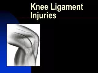
The Knee • Largest joint in the body • Modified hinge joint • One of most vulnerable joints to severe injury of any in the body
Knee Anatomy • Bones • Tibia • Distal to the femur • Major weight bearing bone • Fibula • Not included as a true knee bone • Very little weight bearing • Femur • Longest bone in the body • Major weight bearing • Patella • “floating bone”
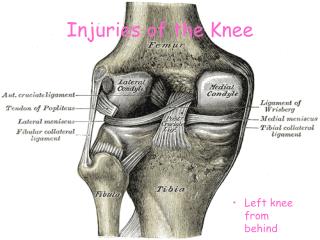
Knee Anatomy • Ligaments • Anterior cruciate ligament (ACL) • “cruciate” means cross • Function of ACL and PCL is to stabilize the knee from front-to-back • Posterior cruciate ligament (PCL) • Medial collateral ligament (MCL) • Lateral collateral ligament (LCL)
ligaments • Posterior cruciate ligament (PCL) • Medial collateral ligament (MCL) • Lateral collateral ligament (LCL)
Knee Anatomy • Cartilage (meniscus) • menisci are horseshoe-shaped shock absorbers that help to both center the knee joint during activity and to minimize the amount of stress on the articular cartilage.
Meniscus • Medial • More often injured than lateral • Often involved medial ligament • C-shaped • Lateral • O-shaped
Knee anatomy • Patellar tendon • Runs from the quadricep muscles, across the patella, and inserts into the tibial tuberosity
Knee Anatomy • Muscles and tendons • Quadriceps-responsible for knee extension • Vastus lateralis • Vastus medialis • Vastus intermedius • Rectus femoris
Posterior Leg • Hamstrings-responsible for knee flexion • Biceps femoris • Semimenbranosis • semitendinosis
Injury prevention • Structural alignment can predispose an athlete to injury
Injury Prevention • Proper strengthening and flexibility of quadriceps, hamstrings, and gastrocnemius muscles
Injury Prevention • Preventative bracing for collateral ligaments
Knee injuries and Conditions • Ligament Injuries • Sprains (ACL, PCL, MCL, LCL) • 1st, 2nd, and 3rd degree • Muscle and tendon injuries • Patellar tendinitis • Bone injuries • Chondromalacia • Patellar dislocations • Other common injuries • Meniscal injuries • Osgood-Schlatter disorder
ACL injuries • Function is to prevent tibia from moving forward on femur • S/S of injury include the athlete feeling disabled, complain of the knee giving way, collapsing, and popping
ACL Injuries • Usually the most serious of all knee injuries • Can hear a pop or snap on injury • Often injured when athlete is changing direction
ACL (cont.) • Can also be injured due to hyperextension • rapid swelling and loss of function • treatment- RICE, knee immobilizer, crutches, follow-up with orthopedist • Almost always require surgical reconstruction if torn
Surgical procedures • Tendon graft • Patellar or hamstring • Allograft • Cadaver tendon
Rehabilitation • 3 Phases of rehab include: • controlling the pain and swelling in the knee • regaining knee motion • beginning to regain muscle strength • Usually minimum 6 months • Conservative treatment for less active people can be non-surgical and focus on all rehab • three components of non-surgical treatment are physical therapy, activity modification, and the use of a brace
PCL injuries • PCL prevents posterior tibial movement on the femur • MOI: bent knee bears full weight, forced hyperflexion, or a blow to the front of the tibia
PCL • Often minimal swelling • Treatment- RICE, refer to physician • Not often surgically repaired • Rehab focuses on strengthening quad muscles
MCL injuries • Usually results from a direct blow to the outside of the knee • Mild sprains result in joint-line point tenderness, minimal swelling, and no joint laxity
MCL • Moderate produces more swelling, discomfort, some loss of function, and some laxity • Severe – produces large amount of laxity
MCL (cont.) • Treat with RICE if mild • Moderate, may need immobilizer, rehab • Moderate to severe could involve the meniscus and/or ACL and may require surgery
LCL injuries • Less common than MCL injuries • Usually occurs due to direct blow to medial side of knee
LCL • Similar s/s except discomfort is on lateral aspect of knee • Focus rehab on lateral thigh muscles and hamstrings
Muscle and tendon injuries • Patellar tendinitis • Characterized by quad weakness and tenderness over patella • Minimal swelling • Called jumper’s knee
Patellar Tendinitis • Pain after activity • Treat with ice, NSAIDs, and restricting activity • Rehab- address flexibility and weakness issues
Bone injuries • Patellar-femoral syndrome • Pain and discomfort around the patella often caused by patellar tracking problems • Causes chondromalacia-the wearing away of the cartilage on the back of the patella
Patellar-femoral syndrome • s/s aching and pain after prolonged sitting, pain when going up or down stairs, athlete feels grinding sensation with flexion/extension
Patellar-femoral syndrome • Treatment involves correcting patellar tracking, strengthening vastus lateralis and medialis, improving flexibility of quads and hamstrings
Bone injuries • Patellar dislocation • Most commonly dislocates laterally • Occurs with bent knee and inward twisting • Noticeable deformity, extreme pain • Call EMS • Physician reduces
Patellar dislocation • Treatment involves immobilization, then rehab to regain mobility and strengthen • Can wear a knee sleeve post-injury to help prevent from happening again
Fractures • Tib-fib fracture • Uncommon, but immediate referral necessary • Many structures involved
Dislocated knee • Extremely rare • Immediate transport
Meniscal injuries Typically occur with a twisting motion or with hyperextension or hyperflexion
s/s-pain over joint line, problems weightbearing, complain of clicking, catching, locking, inability to fully extend or flex, and swelling
Meniscal injuries • Treatment- surgical removal of meniscus (meniscectomy) • More often treated with removal of torn areas only through arthroscopy • Sometimes repair meniscus with sutures or staples • Numerous new methods of repair (i.e. transplants) • Aquatic therapy very useful (non-weight bearing)
Osgood-Schlatter disorder • Inflammation and irritation of the insertion of the patellar tendon (tibial tuberosity) in youth • Repeated stress and activity can cause patella to partially pull away from bone and cause a bump
Osgood-schlatter’s • S/S- pain and discomfort, minimal swelling • Restricted activity recommended • Use pain as a guide for activity level • Ice pre and post activity, NSAIDs
Osgood-schlatter’s • Can try patellar tendon band or pad • Usually improves by age 16 or 17