Download
1 / 33
340 likes | 742 Views
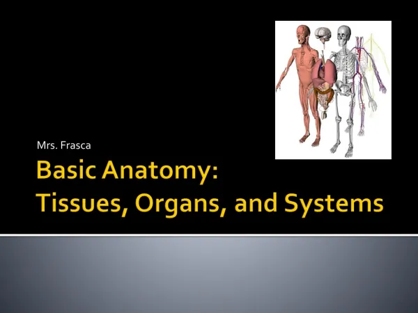
Lecture 12 Body Organs & Tissues. All vertebrates have the same general architecture:. Organization of the Vertebrate Body. Food flows through a long tube from mouth to anus Tube is suspended in coelom , which is divided into Thoracic cavity – Heart and lungs

E N D
All vertebrates have the same general architecture: Organization of the Vertebrate Body • Food flows through a long tube from mouth to anus • Tube is suspended in coelom, which is divided into • Thoracic cavity – Heart and lungs • Abdominal cavity – Stomach and intestines • Body is supported by a skeleton made up of jointed bones • The skull protects the brain • The vertebral column protects the spinal cord
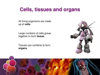
Organs Structures composed of several different tissues grouped into large structural and functional units Organ systems Groups of organs that work together to carry out an important function Levels of organization within the body
There Are 11 Principal Organ Systems • Integumentary system • Skin, hair, nails and sweat glands • Skeletal system • Bones, skull, cartilage, ligaments • Circulatory system • Heart, blood vessels, blood
There Are 11 Principal Organ Systems • Endocrine system • Pituitary, adrenal, ductless glands • Nervous system • Nerve, sense organs, brain, spinal cord • Respiratory system • Lungs, trachea, other air passages
There Are 11 Principal Organ Systems • Immune system • Lymphocytes, macrophages, thymus, lymph nodes • Digestive system • Mouth, esophagus, stomach, intestines • Urinary system • Kidneys, bladder, associated ducts
There Are 11 Principal Organ Systems • Muscular system • Skeletal, cardiac and smooth muscles • Reproductive system • Testes or ovaries • Associated structures
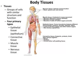
Tissues are collections of cells and cell products that perform specific, limited functions 4 tissue types form all the structures of the human body: Epithelial Tissue Covers external surfaces Lines internal passageways Forms glands Connective Tissue Fills internal spaces Supports other tissues Transports materials Stores energy Muscle Tissue Specialized for contraction Skeletal muscle, heart muscle, and walls of hollow organs Neural Tissue Carries electrical signals from 1 part of the body to another The Body is Made of Four Tissue Types
Epithelium is Protective Tissue • The vertebrate body consists of one tube (digestive tract) suspended into another (body cavity: coelom) • The outside of the body is covered with cells (skin) derived from embryonic ectoderm tissue • The body cavity is lined with cells derived from embryonic mesoderm tissue • The hollow inner core of the digestive tract is lined with cells derived from embryonic endoderm tissue
Epithelia Characteristics • Cellularity (cell junctions) • Polarity (apical and basal surfaces) • Attachment (basal lamina) • Avascularity • Regeneration Functions • Provide physical protection • Control permeability • Provide sensation • Produce specialized secretions (glandular epithelium) Specializations • Move fluids over the epithelium (protection) • Move fluids through the epithelium (permeability) • Produce secretions (protection and messengers)
Epithelial cells are classified into three types according to their shape Classification of Epithelial Cells • Squamous • Cuboidal • Columnar
Simple epithelium Only a single layer thick Found in the lining of the lungs and major body cavities Stratified epithelium Several layers thick Found in the skin Glands Involved in secretion Endocrine glands secretes hormones into the blood Exocrine glands use ducts to secrete sweat, milk, saliva and digestive enzymes out of the body onto epithelial surfaces Three general kinds of epithelial tissue
Connective tissue is derived from the mesoderm Three functional categories 1. Immune system: body defense 2. Skeletal system: body support 3. Blood and fat cells: storage and distribution of substances Connective Tissue Supports the Body
Immune Connective Tissue • Two principal immune cells are • 1. Macrophages • Engulf and digest invading microbes • 2. Lymphocytes • Make antibodies • Or • Attack virus-infected or cancerous cells
Skeletal Connective Tissue • Fibroblasts • The most common kind • Secrete structurally strong proteins such as collagen into spaces between cells • Loose & Dense types • Cartilage • Collagen matrix forms in long parallel arrays along lines of mechanical stress • Found in joint surfaces • Bone • Collagen fibers are coated with calcium phosphate
Storage and Transport Connective Tissue • Includes • Adipose tissue • Accumulates fat • Erythrocytes (RBC) • Transport O2 and CO2 in the blood • The fluid portion of blood is called plasma • Contains nutrients, wastes and antibodies
The distinguishing characteristic of muscle cells is the abundance of contractible protein fibers These microfilaments (myofilaments) are made up of actin and myosin Muscle contraction occurs when actin and myosin slide past each other The vertebrate body possesses three different kinds of muscle cells Muscle Tissue Lets the Body Move • Smooth • Skeletal • Cardiac
Smooth Muscle • Cells are long and spindle-shaped • Each contains a single nucleus • Cellular microfilaments are loosely organized Found in the walls of blood vessels, stomach and intestines Power rhythmic involuntary contractions Sheets of cells
Skeletal Muscle • Produced by fusion of several cells at their ends • This creates a very long muscle fiber that contains all the original nuclei Microfilaments are bunched together into myofibrils • Found in voluntary muscles • Power voluntary contractions Striated
Cardiac Muscle • Composed of chains of single cells, each with its own nucleus • Chains are interconnected, forming a latticework Each heart cell is coupled to its neighbors by gap junctions • Allow electrical signals between cells • Cause orderly pulsation of heart Striated
Nerve tissue is composed of two kinds of cells Neurons Specialized for the transmission of nerve impulses Neuroglial cells Support neurons with nutrients, support and insulation Nerve Tissue Conducts Signals Rapidly • Neuron plasma membranes are rich in ion-selective channels • These maintain a voltage difference between the cell’s interior and exterior • Depolarization is the temporary loss of this voltage difference • It results in a wave of electrical activity, or nerve impulse
Each neuron consists of three parts • 1. Cell body – Contains the nucleus • 2. Dendrites – Bring nerve impulses to the cell • 3. Axon – Carry nerve impulses away from the cell
Skin (Integument) • Consists of three major regions • Epidermis – outermost superficial region • Dermis – middle region • Hypodermis (subcutaneous layer) – deepest region Largest Organ in Body • about 15% of our total weight • Protects underlying tissues and organs • Excretes salts, water, and organic wastes (glands) • Maintains body temperature (insulation and evaporation) • Synthesizes vitamin D3 • Stores lipids • Detectstouch, pressure, pain, and temperature
Skin or Cutaneous Surface Epidermis • 10-30 cells thick (epithelial tissue) • Has no blood vessels • Stratum corneum – Outermost layer • Cells continuously replaced by others from below • Basal layer – Innermost layer Dermis • 15-40 times thicker than the epidermis (loose connective & nerve tissue, + blood vessels) • Provides structural support and nutrients for the epidermis Subcutaneous layer • Fat-rich cells that act as shock absorbers and insulators
Epidermal/Dermal Ridges • Dermal papillae (tiny mounds): • increase the area of basal lamina • strengthen attachment between epidermis and dermis • Epidermal ridges (e.g., fingerprints)
Skin Color • Melanin – yellow to reddish-brown to black pigment, responsible for dark skin colors • Freckles and pigmented moles result from local accumulations of melanin • Melanin protects skin from sun damage (UV radiation) • Carotene – yellow to orange pigment • Most obvious in the palms and soles of the feet • Can be converted to vitamin A • Hemoglobin – reddish pigment responsible for the pinkish hue of the skin Three pigments contribute to skin color
Cutaneous Glands Sweat (Sudoriferous) Glands • Different types prevent overheating of the body; secrete cerumen and milk • Eccrine sweat glands – found in palms, soles of the feet, and forehead • Apocrine sweat glands – found in axillary and anogenital areas • Ceruminous glands – modified apocrine glands in external ear canal that secrete cerumen • Mammary glands – specialized sweat glands that secrete milk Sebaceous Glands • Simple alveolar glands found all over the body • Soften skin when stimulated by hormones • Secrete an oily secretion called sebum • Acne occurs when these glands become blocked or infected
Basal Cell & Squamous Cell Carcinomas • Basal Cell Carcinoma • Least malignant and most common skin cancer • Stratum basale cells proliferate and invade the dermis and hypodermis • Slow growing and do not often metastasize • Can be cured by surgical excision in 99% of the cases • Squamous Cell Carcinoma • Arises from keratinocytes of stratum spinosum • Arise most often on scalp, ears, and lower lip • Grows rapidly and metastasizes if not removed • Prognosis is good if treated by radiation therapy or removed surgically
Melanoma • Cancer of melanocytes is the most dangerous type of skin cancer because it is: • Highly metastatic • Resistant to chemotherapy • Melanomas have the following characteristics (ABCD rule) • A:Asymmetry; the two sides of the pigmented area do not match • B:Border is irregular and exhibits indentations • C:Color (pigmented area) is black, brown, tan, and sometimes red or blue • D:Diameter is larger than 6 mm (size of a pencil eraser) • Treated by wide surgical excision accompanied by immunotherapy • Chance of survival is poor if the lesion is over 4 mm thick
Burns • First-degree – only the epidermis is damaged • Symptoms include localized redness, swelling, and pain • Second-degree – epidermis and upper regions of dermis are damaged • Symptoms mimic first degree burns, but blisters also appear • Third-degree – entire thickness of the skin is damaged • Burned area appears gray-white, cherry red, or black; there is no initial edema or pain (since nerve endings are destroyed) • Destruction of proteins of the skin • chemicals, electricity, heat • Problems that result • shock due to water, plasma and plasma protein loss • circulatory & kidney problems from loss of plasma • bacterial infection
Rule of Nines • Estimates the severity of burns • Burns considered critical if: • Over 25% of the body has second-degree burns • Over 10% of the body has third-degree burns • There are third-degree burns on face, hands, or feet Figure 5.8a
Other Epithelial Surfaces • Serous – moist membranes found in closed ventral body cavity • Mucous – lines body cavities open to the exterior (e.g., digestive and respiratory tracts)