
Hemodynamic Dysfunction & Related Conditions
E N D
Presentation Transcript
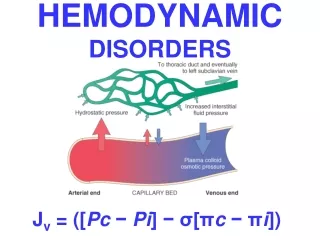
Hemodynamic Dysfunction Edema: - This is an abnormal accumulation of fluid in interstitial tissue spaces or body cavities. A. Causes of edema 1. Increased hydrostatic pressure is exemplified by CHF. 2. Increased capillary permeability occurs in inflammation or with injury to capillary endothelium 3. Decreased oncotic pressure is from hypo-albuminemiacaused by: a. Increased loss of protein, for example, by renal loss in the nephrotic syndrome b. Decreased production of albumin in cirrhosis of the liver 4. Increased sodium retention can occur as either a primary or secondary phenomenon. a. Primary sodium retention, associated with renal disorders b. Secondary sodium retention, such as occurs in CHF
5. Blockage of lymphatics results in lymphedema. Note: A transudate: occurs with volume or pressure overload, or under conditions of reduced plasma protein; typically it is protein_poor with specific gravity less than 1.012 An exudate it is protein rich with specific gravity greater than 1.020, it occurs due to increased vascular permeability in inflammation.
Hemorrhage A. general considerations1. Hemorrhage is the extravasation of blood from the vasculature into surrounding tissues, a hollow organ or body cavity, or to the outside.2. Hemorrhage is most often caused by trauma. 3. Hemorrhagic diatheses increased tendency to hemorrhage. B. Hematoma. This localized hemorrhage occurs within a tissue or organ, can be relatively in significant (e.g., bruise) or can be massive bleeding and cause death (e.g., massive retroperitoneal hematoma).
C. Hemothorax, hemopericardium, hemoperitoneum, and hemarthrosis. Hemorrhage may occur in the pleural cavity, pericardial sac, peritoneal cavity, or a synovial space, respectively.D. Petechial hemorrhages, petechiae, or purpura. These small, punctate hemorrhages occur in the skin, mucous membranes, or serosal surfaces.E. ecchymosis. This diffuse hemorrhage is usually in skin and subcutaneous tissue.
Hyperemia: - a localized increase in the volume of blood in capillaries and small vessels. A. active hyperemia. The cause is localized arteriolar dilation (e.g., blushing, inflammation).B. Passive congestion (passive hyperemia). The cause is obstructed venous return or increased back pressure from congestive heart failure (CHF).1. Acute passive congestion occurs in shock, acute inflammation, or sudden right-sided heart failure.2. Chronic passive congestiona. chronic passive congestion of the lung is caused most often by left-sided heart failure or mitral stenosis.
Infarction: - A. Definition. Infarction is localized area of ischemic cell necrosis in a living organ or tissue, result from sudden reduction or cessation of its arterial blood supply or occasionally its venous drainage. B. Anemic infarcts1. These infarcts are white or pale infarcts.2. They are usually caused by arterial occlusions in the heart, spleen, and kidney.
C. Hemorrhagic infarcts1. These infarcts are red infarcts, in which red cells ooze into the necrotic area.2. They occur characteristically in the lung and gastrointestinal tract as the result of arterial occlusion. occurs from the no obstructed portion of the vasculature.3. They can also be caused by venous occlusion. This is an important contribution to infarcts associated with volvulus, incarcerated hernias, and postoperative adhesions.
Thrombosis: - intravascular coagulation of blood, often causing significant interruption of blood flow. Pathologically predisposed by many conditions, including venous stasis, usually from immobilization; CHFpolycythemia sickle cell disease visceral malignancies and the use of oral contraceptives, especially in association with cigarette smoking.
Thrombogenesis. This process results from the interaction of 1-platelets, 2-damaged endothelial cells(vascular wall), and 3-the coagulation cascade. • 1. Platelets a. Platelet functions (1) Maintain the physical integrity of the vascular endothelium (2) Participate in endothelial repair through the contribution of platelet-derivedgrowth factor (PDGF) (3) Form platelet plugs (4) Promote the coagulation cascade through the platelet phospholipid complex