Download
1 / 32
340 likes | 460 Views
In this study, we explore the structural basis for mismatch replication errors in DNA polymerases, focusing on the high-fidelity Bacillus DNA polymerase I fragment (BF). We employ crystallographic techniques to capture and analyze the twelve potential mismatches at the polymerase's active site. Our findings reveal distinct structural alterations associated with each mismatch, influencing polymerase stalling and accuracy. The results highlight the critical interactions within the catalytic site and illustrate the diverse impacts of mismatches on DNA replication fidelity.

E N D
Structures of Mismatch Replication Errors Observed in a DNA Polymerase Sean J. Johnson and Lorena S. Beese Biochemistry 4000 Tracy Wang
High-fidelity DNA polymerases maintain replication accuracy through: • Select for correct base pairing, while strongly discriminating against mismatched bases ( prior to covalent incorporation) • Stalling of the polymerases, thereby favoring subsequent mismatch excision (after incorporation)
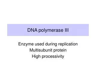
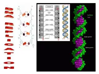
Klenow Fragment • DNA polymerase I • Highly processive • Fingers, Thumb and Palm
Taq polymerase • Closed conformation • dNTP bound • Open conformation • No incoming nt.
Model system • Use the thermophilic Bacillus DNA polymerase I fragment ( BF). • BF is a high-fidelity Family A polymerase. • Structural homology to the Klenow Fragement of E.coli (KF) and T. aquaticus (Taq) polymerases.
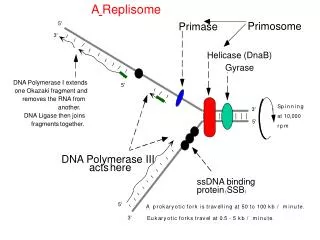
Model system • Insertion site (n position ) • Catalytic site • Preinsertion site • Postinsertion site (n-1 position ) • DNA duplex binding region (n-2,…, n-5 positions )
Replication mechanism • Transfer of the n template base from the pre-insertion site to the insertion site: - open to closed conformation - hinge-bending motion • Base pairs with an incoming base, covalent incorporation takes place. • Newly synthesized base moves into the post-insertion site. • The DNA in the duplex binding region translocates by one base pair. • The next template base moves into the pre-insertion site.
Cognate G•C base pair bound at the postinsertion(n-1) site. • Hydrogen bond between Asp830 and the 3’ primer terminus. • Arg615 and Gln797 interact with hydrogen bond acceptor atom located in the DNA minor groove.
Objective • To obtain and solve the structure of all 12 possible mismatches captured at the active site of a DNA polymerase. • To understand the mechanisms that lead to mismatch-induced stalling of the polymerase.
Crystallographic capture of mismatches • Crystals with mismatches were obtained either by enzymatic incorporation in the crystals (E), or by cocrystallization of DNA duplexes that contain a mismatch at the primer terminus (C). • In the presence of Mg2+, accurate DNA replication is obtained. • In the presence of Mn2+, enzymatic incorporation of mismatches is allowed. • E: use mutagenic reaction conditions (MgSO4 MnSO4 ) • C: BF cocrystallized with DNA substrates
Results • 12 possible covalently incorporated DNA mismatches. • Six are placed at the post-insertion site and are well ordered (G•T, T•G, T•T, C•T, A•G, G•G). • Three are placed at the post-insertion site but are too disordered for interpretation (T•C, A•C, C•A). • Three are frayed (A•A, C•C, G•A). Primer strand is at the insertion site Template strand is at the pre-insertion site
Results • Structure of each mismatch is distinct. • Some structures had been previously observed, others do not. • The type and degree of disruptions vary depending on the identity of the mismatch.
Mismatch-induced disruptions at the active site • Displacement of the template strand • Repositioning of Tyr714 • Block the template pre-insertion site due to the rearrangement • B form DNA at the active site rather than A form • The catalytic site is undisturbed
G adopts a conformation similar to that of a cognate base intact catalytic site • Wobble conformation, T is positioned toward DNA major groove • Minor groove interaction with Gln797 is lost displacement of template strand • Primer base G rotates 180 into a syn conformation, template base remains in an anti conformation helical width is closer to a Watson-Crick base pair • More closely resembles the G•T complex • Template strand is displaced • N3 is moved from the minor groove interaction between Arg615 and N3 is lost
T•C, A•C : disordered mismatches • The disorder is confined primarily to the template strand • Disorder on the primer strand is localized to the primer base • A mixture of A and B forms of DNA can be discerned in the DBR. • Protein adopts a distorted open conformation in which the pre-insertion site is blocked.
The primer terminus is displaced • Interaction between Asp830 and the primer 3’ hydroxyl at the catalytic site is disrupted • The DNA backbone of the template strand is undisturbed
wobble conformation. • The primer base lifts up into the DNA major groove, and the template base rotates slightly toward the minor groove. • Shift of the 3’ hydroxyl breaking the hydrogen bond with Asp830 disrupting assembly of the catalytic site. • Not a wobble conformation. • Base pair directly opposite each other • Significant opening in the minor groove • Bridging water molecule
Displacement of the template strand. • Blocking of the pre-insertion site. • Displacement of the primer strand. • Disruption of the catalytic site.
Both bases maintain an anti conformation increase of the helical width extensive movement of both template and primer strands. • Altering the interaction with Asp830, but without breaking it. • Wobble conformation. • An additional water-mediated H-bond. • Template is displaced • Interaction between Arg615 and the primer base is lost • Sugar ring of T is disordered primer terminus is displaced, but interaction between Asp830 and primer 3’ hydroxyl may be retained to some degree.
Structure is frayed. • The primer base is bound at the insertion site. • The template base is bound at the pre-insertion site.
A•A, C•C, G•A: frayed mismatches. • Do not bind in the post-insertion site. • Tyr714 blocks access of the template base to the insertion site. • Tyr714 also prevents the primer base from stacking to the DNA helix. Primer base stacks against Phe710. • Catalytic site is completely disrupted.
Mismatch extensions • By transferring BF.DNA cocrystals containing a mismatch at the 3’ primer terminus into reaction buffers containing nucleotides complementary to the template strand. • By soaking in different combinations of nucleotides in the presence of Mg2+ to control the final location of the mismatch
Results • The A•G, T•T, T•G, and C•C mismatches failed to extend. • The G•T, C•T, and G•G mismatches were all successfully extended. • For the T•C mismatch, DNA is too disordered to permit interpretation.
G•T Extension • At n-2 position: wobble • At n-2 and n-3: disrupt A to B form transition, similar to the disruptions observed when G•T is at n-1 disrupted active site • At n-4: partially restores the normal DNA structure mixture of both a disrupted and an undisrupted active site • At n-3 and n-4 : the wobble inverts inconsistent with the interbase H-bonding geometry associated with the major tautomeric form of nt. tautomeric shift or ionization of the mismatch • At n-6: fully restores the DNA conformation
C•T Extension • Extended in a single round of replication • At n-2 position: fully restores the catalytic site.
Summary • The high-resolution structures of all 12 possible covalently incorporated DNA mismatches at the active site of the BF polymerase were obtained. • The mismatch-induced disruptions can be divided into four broad categories. • Disruption of template strand and pre-insertion site. • Disruption of primer strand and assembly of catalytic site. • Disruption of template and primer strands. • Fraying of DNA at insertion site. • The polymerase retains a short-term memory of the mismatch incorporation event.
Discussion • Category of mismatch-induced disruptions differences in mismatch extension efficiencies ? • G•T: disruption may account for the reduced extension efficiency ten-fold to a thousand-fold. G•G: more than 100-fold slower than that of G•T. • C•T: can be extended readily due to the flexibility of DNA. • A•G: efficiencies are reduced by up to million-fold.
Discussion • A heteroduplex can adjust its size in order to translocating through the DBR. • The structural adaptations are confined primarily to the DNA, with some local protein side chain motions. • Exonucleolytic excision of mismatches requires dissociation of the heteroduplex from the polymerase: A number of the structures show diminished interactions. • Some mismatch conformations have not been previously described in a DNA heteroduplex. - Structural adaptations are not confined to simple conformational rearrangements. • Each equivalent mismatch interacts in a unique manner with the protein. (e.g. G•T, T•G)
Future work • Basis for future biochemical and structural studies. • Study the structural changes of a given mismatch under different conditions (pH, sequence context). • To see if these mechanisms can be applied to other DNA polymerases and even RNA polymerases.
References • Johnson, S. J. and Beese, L. B. Cell 2004, 116, 803-816.