Download
1 / 20
881 likes | 4.68k Views
Serum Creatine kinase. Description. Creatine kinase (CK), also known as creatine phosphokinase (CPK) or phospho-creatine kinase is an enzyme ( EC 2.7.3.2 ) expressed by various tissues and cell types. Function.

E N D
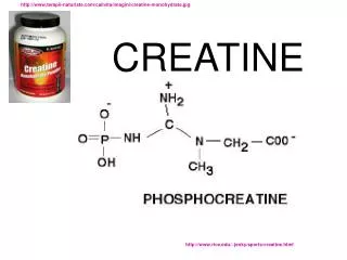
Description • Creatine kinase (CK), also known as creatine phosphokinase (CPK) or phospho-creatine kinase is an enzyme (EC2.7.3.2) expressed by various tissues and cell types.
Function • CK catalyses the conversion of creatine and consumes adenosine triphosphate (ATP) to create phosphocreatine (PCr) and adenosine diphosphate (ADP). • This CK enzyme reaction is reversible, such that also ATP can be generated from PCr and ADP.
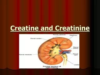
Tissue Origin • It is In tissues and cells that consume ATP rapidly, especially skeletal muscle, but also brain, of and cardiac muscle. • phosphocreatine serves as an energy reservoir for the rapid buffering and regeneration of ATP in situ.
Isozymes • Isozymes are enzymes that catalyze the same reaction. However, they do not have the same physical properties because of genetically determined differences in amino acid sequence. • Isoenzymes may contain different numbers of charged amino acids and may, therefore, be separated from each other by electrophoresis. • Different organs frequently contain characteristic proportions of different isoenzymes. The pattern of isoenzymes found in the plasma may therefore, serve as a means of identifying the site of tissue damage.
CK isoenzymes In the cells, the "cytosolic" CK enzymes consists of two subunits, which can be either B (brain type) or M (muscle type). There are, therefore, three different isoenzymes: • CK1=CK-BB=brain type • CK2=CK-MB=cardiac type • CK3=CK-MM=muscle type
Skeletal muscle expresses CK-MM (98%) and low levels of CK-MB (1%). • The myocardium (heart muscle), in contrast, expresses CK-MM at 70% and CK-MB at 25–30%. • CK-BB is expressed in all tissues at low levels and has little clinical relevance.
While mitochondrial creatine kinase is directly involved in formation of phospho-creatine from mitochondrial ATP, cytosolic CK regenerate ATP form ADP.
Diagnostic Use Clinically, creatine kinase is assayed in blood tests as a marker of: • Myocardial infarction (heart attack), • Muscular dystrophy,(severe muscle breakdown), • Central nervous system lesions.
In myocardial infarction it begins to rise in 4-8 hours • reachs its maximum level in 12-24 hours • returns to normal level after 2 or 3 days. Normal level at 37 C: 24 -190 u/L in male. 24- 170 u/L in female.
ADP = Adenosine 5'-Diphosphate ATP = Adenosine 5'-Triphosphate G-6-PDH = Glucose-6-Phosphate Dehydrogenase ß-NADP = ß-Nicotinamide Adenine Dinucleotide Phosphate, Oxidized Form ß-NADPH =ß-Nicotinamide Adenine Dinucleotide Phosphate, Reduced Form 6-PG = 6-Phospho-D-Gluconate CONDITIONS:T = 30°C, A 340 nm. Specimen: Serum, heparinised plasma and Edta plasma.
Electrophoresis • Electrophoresis is the migration of charged molecules in a media upon application of an electric field. • The rate of migration depends on the charge on the molecule, its molecular mass and the strength of the electric field. • Usually, used for the separation of proteins.
The most commonly used matrix is agarose. • Agarose is used mostly for separation of larger macromolecules, including proteins and their complexes. • Proteins can be visualized after electrophoresis by treating the gelwith a stain such as Coomassie blue. • Note: Proteins in alkaline buffer carry negative charge and migrates to anode (positive electrode).
Different CK isoenzymes pattern can be detected by electrophoresis.
SDS-PAGE • Catalase, cytochrome C, a-lactalbumin • Hemoglobin, Cytochrome C, a-lactalbumin • BSA, cytochrome C, a-lactalbumin • Hemoglobin, myoglobin, a-lactalbumin • Ferritin, cytochrome C, a-lactalbumin • Ferritin, myoglobin, a-lactalbumin 1 2 3 4 5 6 lighter
Agarose Gel Stained with ethidium bromide (EtBR) to Visualize the DNA slots where DNA is loaded 1000 bp 700 bp 600 bp 500 bp Screening PCR products to test for the presence of specific DNA sequences molecular weight markers correct PCR product molecular weight markers