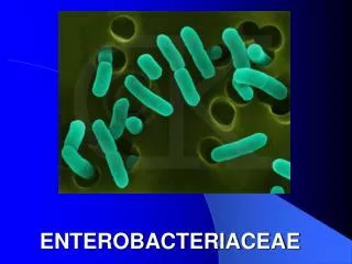
ENTEROBACTERIACEAE
Learn about the characteristics of Enterobacteriaceae, including gram-negative bacilli, ability to ferment glucose and reduce nitrates, and their clinical significance in causing meningitis, sepsis, diarrhea, and UTIs.


ENTEROBACTERIACEAE
E N D
Presentation Transcript
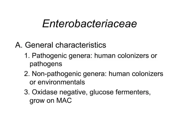
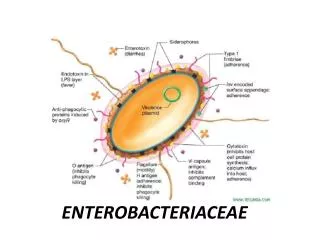
Characteristics of the Enterobacteriaceae • Gram-negative bacilli • Gram-negative bacilli • Ferment glucose • Reduce nitrates to nitrites • Oxidasenegative • Catalase positive (except Sh.disentry type 1) • All except Klebsiella, Shigella and Yersinia are motile by peritrichous flagella
Grow readily on • MacConkey (MAC) agar • Eosin methylene blue (EMB) agar • Grow readily at 35oC except Yersinia (25o-30oC) • Do not form spores • Natural Habitat: • Environment (soil, water, and plants) • Intestines of humans and animals
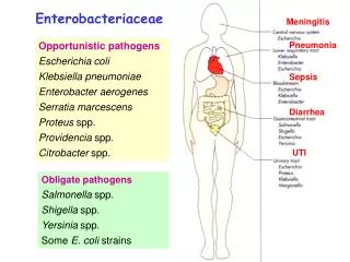
Enterobacteriaceae Meningitis Opportunistic pathogens Escherichia coli Klebsiella pneumoniae Enterobacter aerogenes Serratia marcescens Proteus spp. Providencia spp. Citrobacter spp. Pneumonia Sepsis Diarrhea UTI Obligate pathogens Salmonella spp. Shigella spp. Yersinia spp. Some E. coli strains
Classification of Enterobacteriaceae: - Based on lactose fermentation • LF • LLF • NLF
Classification of Enterobacteriaceae: • Very large number of organisms in Family Enterobacteriaceae, species are grouped into - TRIBES • Tribe 1 – Escherichieae • Tribe 2 – Klebsiellae • Tribe 3: Proteae • Tribe 4 : Yersiieae • Tribe 5 : Erwinieae • Within each Tribe, species are further subgrouped under genera
Major Genera of family Enterobacteriaceae: Escherichia Shigella Salmonella Yersinia Klebsiella Proteus
Antigenic Factors of Enterobactericeae: • Ability to colonize, adhere, produce various toxins and invade tissues • Some possess plasmids that may mediate resistance to antibiotics • Imp antigens used to identify the organisms: • O antigen – somatic, cell wall, heat-stable Ag • H antigen – flagellar, heat labile Ag • K antigen – capsular, heat-labile Ag
Clinical Significance of Enterics: • Ubiquitous in nature • Except for Salmonella, Shigella and Yersinia, most are present in the intestinal tract of animals and humans as commensal flora; “fecal coliforms” • Some live in water, soil and sewage
Clinical Significance of Enterics: • Based on clinical infections produced, enterics are divided into two categories: • Opportunistic pathogens – normally part of the usual intestinal flora, that may produce infection outside the intestine • Primary intestinal pathogens – Salmonella, Shigella, and Yersinia sp.
Enterobacteriaceae: Modes of Infection • Contaminated food and water (Salmonella spp., Shigella spp., Yersiniaenterocolitica, Escherichia coli O157:H7) • 2. Endogenous infection (urinary tract infection, primary bacterial peritonitis, abdominal abscess) • 3. Abnormal host colonization (nosocomial pneumonia) • Transfer between debilitated patients • 4. Insect (flea) vector: unique for Yersiniapestis
Urinary tract infection: Escherichia coli, Klebsiellapneumoniae, Enterobacter spp., and Proteus mirabilis • Pneumonia: Klebsiellapneumoniae; Enterobacter spp., • Wound Infection: Escherichia coli, Enterobacter spp., Klebsiellapneumoniae, and Proteus mirabilis • 4. Bacteremia: Escherichia coli, Enterobacter spp., Klebsiellapneumoniae, and Proteus mirabilis
Enterobacteriaceae: Intestinal Infection • Escherichia coli • Shigella • Salmonella • Yersinia enterocolitica
Identification of Enterobacteriaceae: • IMViC Test • Indole, Methyl Red, Voges-Prosakaur, Citrate (IMViC) Tests: • The IMViC series of reactions allows for the differentiation of the various members of family Enterobacteriaceae.
A) Indole –Positive, B) MR –Positive C) VP- Positive, D) Citrate- Negative
Methyl Red-Voges Proskauer (MR-VP) Tests Principle Glucose Acidic pathway Or Neutral pathway Acety methyl carbinol (ACETOIN) Mixed acids pH less than 4.4 Barrit’s A Barrit;s B Methyl Red indicator VP positive Klebsiella MR positive E. coli Pink color Red color
MR/VP test Voges-Proskauer test Methyl Red test • Pink: Positive VP (Klebsiella) • Red: Positive MR (E. coli) • No pink: Negative VP (E. coli) • Yellow or orange: Negative MR (Klebsiella)
TSI Reactions of the Enterobacteriaceae • A/A + g = acid/acid plus gas (CO2) • A/A = acid/acid • A/A + g, H2S = acid/acid plus gas, H2S • Alk/A = alkaline/acid • Alk/A + g = alkaline/acid plus gas • Alk/A + g, H2S = alkaline/acid plus gas, H2S • Alk/A + g, H2S (w) = alkaline/acid plus gas, H2S (weak)
A/A + g • Escherichia coli • Klebsiella A/A + gas, H2S • Citrobacter freundii • Proteus vulgaris • Non-lactose, sucrose fermenter
Alk/A • Shigella • Providencia Alk/A + g • Salmonella serotype Paratyphi A Alk/A + g, H2S • Salmonella • Proteus mirabilis • Edwardsiella tarda
Lactose fermenting colonies Non lactose fermenting colonies
Escherichia coli • Most significant species in the genus • Important potential pathogen in humans • Common commensal in the intestine • Pink (lactose positive) colony with surrounding pink area on MacConkey
Ferments glucose, lactose, trehalose, & xylose • Usually motile • Positive indole and methyl red tests • Does NOT produce H2S • Simmons citrate negative • Voges-Proskauer test negative
Pathogenesis and clinical diseases Sepsis In people with inadequate host defenses, e.g. the newborns. Usually originates from UT or GI infections. Some infections may be endogenous. Meningitis E. coli (particularly K1 strains) and S. agalactiae are the leading causes of meningitis in infants. Bacteremia
Urinary tract infection E. coli is the most common cause of urinary tract infection. Community- vs. hospital-acquired UT infection Most infections originate from colon; the bacteria contaminate the urethra, ascend into the bladder, and may migrate into the kidney or prostate. Symptoms: urinary frequency, dysuria, hematuria, and pyuria. Can result in bacteremia and sepsis. UropathogenicE. coli strains produce P (Pyelonephritis-associated) pili, which is associated with renal colonization and may induce protective immunity, and hemolysinHlyA.
Infections caused by E.coli: • Meningitis, gastrointestinal, urinary tract, wound, and bacteremia • Gastrointestinal Infections: • - diarrhoegenic E. coli • Enteropathogenic (EPEC) • Enterotoxigenic E. coli (ETEC) • Enteroinvasive E. coli (EIEC ) • Enterohemorrhagic E. coli (EHEC) • Enteroaggregative E. coli (EAEC)
Enteropathogenic (EPEC) • Primarily in infants and children; • Outbreaks in hospital nurseries and day care centers • Stool has mucous but not blood • Identified by serotyping
Enteropathogenic E.coli Destruction of surface microvilli • Fever • Diarrhea • Vomiting • Nausea • Non-bloody stools Gut lumen
Enterotoxigenic E. coli • Diarrhea resembling cholera • Travelers diarrhea
Enterotoxigenic E. coli • Heat labile toxin • Like cholera toxin • Adenyl cyclase activated • Cyclic AMP • Secretion water/ions • Heat stable toxin • Guanylate cyclase activated • cyclic GMP • uptake water/ions
Enteroinvasive E. coli (EIEC ) • Dysentery - Resembles shigellosis
Transmission electron micrograph Enterohemorrhagic E. coli • Usually O157:H7 Flagella
O157:H7transmitted- through meat products or sewage-contaminated vegetables • Hemorrhagic diarrhea • Bloody, copious diarrhea • Few leukocytes • Afebrile • Hemolytic-uremic syndrome (HUS) • Hemolytic anemia • Thrombocytopenia (low platelets) • Kidney failure
Enterohemorrhagic E. coli • Vero toxin- “shiga-like” • Hemolysins Enteroaggregative (EaggEC) Cause diarrhea by adhering to the mucosal surface of the intestine; watery diarrhea; symptoms may persist for over two weeks
Urinary Tract Infections • E. coli is most common cause of UTI and pyelo-nephritis in humans • Usually originate in the large intestine • Able to adhere to epithelial cells in the urinary tract
Septicemia & Meningitis • Most common causes of septicemia and meningitis among neonates • Acquired in the birth canal before or during delivery • E. coli also causes bacteremia in adults, secondary to genitourinary tract infection or a gastrointestinal source
Klebsiella, Enterobacter • Usually found in intestinal tract • Wide variety of infections, primarily pneumonia, wound, and UTI • General characteristics: • Non-motile • Simmons citrate positive • H2S negative, Weakly urease positive • MR negative; VP positive
Usually found in Gastro-intestinal tract • K. pneumoniae is most commonly isolated species • Possesses a polysaccharide capsule - which protects against phagocytosis and antibiotics • Makes the colonies moist and mucoid • Frequent cause of nosocomial pneumonia
Significant biochemical reactions • Lactose positive • Most are urease positive • Non-motile
Nosocomial pneumonia:Spread by health care personnel and equipment • Frequently caused by K. pneumoniae • Often seen in middle-aged males who abuse alcohol • Difficult to diagnose due to commensals in sputum
Proteus, Morganella & Providencia species • All are normal intestinal flora • Opportunistic pathogens • Deaminate phenylalanine • Non lactose fermenters
Proteus species • P. mirabilis and P. vulgaris are widely recognized human pathogens • Isolated from urine, wounds, and ear and bacteremic infections • Both produce swarming colonies and have a distinctive “burned chocolate” odor • Both are strongly urease positive • Both are phenylalanine deaminase positive
Laboratory Diagnosis of Enterics • Specimen collection: • Specimens collected and transported in Cary-Blair, Amies, or Stuart media Isolation and Identification • Site of origin must be considered • Enterics from sterile body sites are highly significant • Routinely cultured from stool
Media for Isolation and Identification of Enterics: • Blood agar and a selective/differential medium such as MacConkey • On MacConkey, lactose positive are pink; lactose negative are clear and colorless
For stool, highly selective media, such as Hektoen Enteric (HE), XLD, or SS is used along with MacConkey agar • Identification: All enterics are • Oxidase negative • Ferment glucose • Reduce nitrates to nitrites