Cell Communication
Cell Communication. Chapter 11. Signal Transduction Pathway. Series of steps/ reactions in a cell that are caused by a signal on the cell’s surface that triggers a specific cellular response For example: sugar molecule on taste bud. Evolutionary Link.

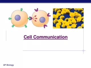
Cell Communication
E N D
Presentation Transcript
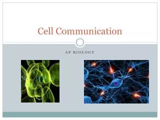
Cell Communication Chapter 11
Signal Transduction Pathway • Series of steps/ reactions in a cell that are caused by a signal on the cell’s surface that triggers a specific cellular response • For example: sugar molecule on taste bud
Evolutionary Link • Signal transduction pathways are very similar between prokaryotes and eukaryotes • This shows that this type of cell signaling must have evolved very early on in evolutionary history • Yeast cell mate identification (a and a) • Bacterial cell protection when food is scarce
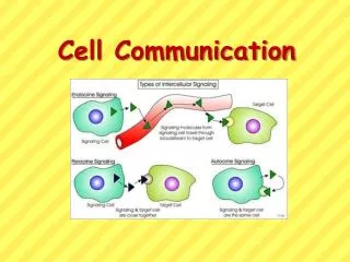
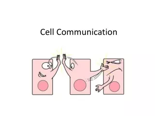
Types of signaling • Local signaling – occurs through direct contact or by signaling molecules • Long distance signaling – occurs through hormones
Local Signaling – Direct • Direct contact • Celljunctions (gap junctions in animals or plasmodesmata in plants) • Cell to Cell recognition (glycoproteins—wewillseethisis the immune system)
Local Signaling – Signaling Molecules • Signaling molecules are secreted by one cell and received by a target cell • Ex – paracrine signaling: secreting cells discharge local regulator molecules to nearby target cells (like growth factors to tell a cell to grow and divide) • Ex – synaptic signaling: nerve cells release neurotransmitters across a synapse, stimulating the target cell
Long Distance - Hormones • Specialized cells (endocrine cells) secrete hormones into vessels; hormones travel through the blood stream to target cells in the body • Ex – insulin from the pancreas targets liver, muscle, and fat to take up glucose and store it as glycogen • Plant hormones can move through vessels or through cells or even through the air as gas • Ex- Ethylene given off by plants for ripening fruit passes through cell wall as a gas
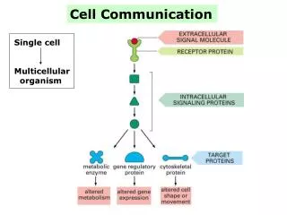
3 stages of cell signaling • Overview: when a cell encounters a signaling molecule is must be 1) recognized by a receptor protein, 2) the signal (info it carries) must be changed into another form inside the cell (transduced), before 3) the cell can respond. • Signaling molecule = ligand • Ligand : a molecule that specifically binds to another molecule (usually a larger one) and usually results in a shape change to the receptor protein
3 stages of cell signaling – stage 1 • 1) Reception: signaling molecule from outside the cell binds to a receptor protein on the cell’s surface • Lock and key (SUPER SPECIFIC) = ligand causes change in shape/receiver = initial transduction • If ligand is hydrophobic (testosterone), the receptor is probably inside the cell • If the ligand is hydrophilic, the receptor is outside the membrane such as: • G-protein-coupled receptors and receptor tyrosine kinases • Ion Channel Receptors – regulate flow of ions
G-protein-coupled receptors Ligand binds to receptor causing shape change in G-protein-coupled receptor G protein is able to attach using GTP for energy (leaving GDP) Activated G protein binds to an enzyme causing ONE cellular response Return to original shapes
What do G proteins do for me? G proteins are used in embryonic development, vision, and smell and are used in many diverse organisms in a similar way Bacteria can interfere with the function of G proteins/receptors – cholera, pertussis (whooping cough), botulism Up to 60% of the medicines we use today influence the G-protein pathway
Receptor Tyrosine Kinase Signal molecules bind to receptors Receptor molecules move together A phosphate from ATP is added to each tyrosine Results in MULTIPLE (10 or more) cellular responses
Ion Channel Receptors Ion channel is closed without the ligand present Binding of the ligand to the receptor on the ion channel opens the gate for ions to flow through from high to low concentration Cause a change in the concentration gradient, triggers cell response Removal of the ligand causes gate to close, preventing ions from moving, thus the cellular response stops AKA – ligand-gated ion channels Used in the nervous system
#2 - Transduction • 2) Transduction: a step or steps to bring about a specific cell response like a shape change of a protein or gene activation • Signal transduction pathway is usually more than one step = CASCADE • Can amplify a signal AND help the cell regulate its processes • Protein Kinase: an enzyme that transfers a P group from ATP to an inactive protein (phosphorylates this protein) • Protein Phosphatase: enzyme that removes a P group (dephosphorylate) from the active protein to turn OFF the signal transduction pathway
#2 - Transduction • Inactive protein kinases are phosphorylated and activated with the help of the previously active protein • Ultimately aid in phosphorylation and activation of a final protein
A Typical Pathway to Cell Division • For normal cell division, each of these proteins have to be activated starting at #10 through #1 to result in a “blast off” into mitosis
Where might errors occur in the transduction pathway? • Cancer can result from one or more of these proteins being active too long , sending the “divide” signal too much, resulting in mitosis that occurs too often • sis – growth factor • erb – receptor protein • src – receptor kinase (activated by a virus) • ras – G protein (activated by a point mutation)
Second Messengers • Small, non-protein, water-soluble molecules or ions that move through diffusion unlike an extracellular ligand attaching to the membrane receptor(first messenger) • Examples are cyclic AMP (cAMP) and calcium ions
Cyclic AMP (cAMP) • cAMP comes from ATP breaking into 2 P + cAMP • Triggered by epinephrine (the ligand/1st messenger) • G protein activates adenylylcyclase • Adenylylcyclase catalyzes the reaction of ATP cAMP + 2P • cAMP can broadcast to entire cytoplasm and activate Protein Kinase A • Will continue to make more cAMP and activate more Protein Kinase A until inhibitors are activated • See cholera example in the book
Calcium ions • Used in muscle contraction, nervous system, cell division among others • Cell pump Ca2+ out of cell, into ER and into mitochondria to keep cytosol concentrations low • During signal transduction pathway, Ca2+ is released from ER and Ca2+ levels rise in cytosol • Ca2+ used as a “third messenger” here but still called second messenger
#3 - Response • 3) Response: cell response is activated (usually by turning genes on or off) • Nuclear response - impact transcription • Cytoplasmic response - impact activity of proteins and impact shape of cell (yeast cells finding mates)
3 stages of cell signaling • REVIEW DIAGRAM
Example of Signal Transduction Pathway • Apoptosis is programmed cell suicide • A normal part of vertebrate development • Nervous system • Hands/feet
Signal for Apoptosis • Signal from outside the cell could be a “cell death” signal sent from a neighboring cell • Signal from inside the cell could come from the • nucleus due to irreparable damage to the DNA • ER due to too many misfolded proteins
A Cellular Response :Apoptosis • Cell is broken down and digested • DNA is chopped • Organelles are fragmented • Cell shrinks • Blebbing occurs (lobes form) • Cell part are packaged in vesicles and digested by scavenger cells = NO TRACE OF CELL REMAINS • Protects nearby cells from damage from dying cell • Cascade of “suicide” proteins
When apoptosis goes wrong… • Too much apoptosis can lead to degenerative diseases such as Parkinson’s or Alzheimer’s • Too little apoptosis can lead to cancer