Download

1 / 26
380 likes | 1.44k Views
URINARY BLADDER & BULBOURETHRAL GLAND. By: MURSHIDA BT SHAHUL HAMEED D11A019 NURUL SYUHADAH BT RIBUAN D11A032 NUR NABILA BT MOHD ROZAINI D11A027

E N D
URINARY BLADDER & BULBOURETHRAL GLAND By: MURSHIDA BT SHAHUL HAMEED D11A019 NURUL SYUHADAH BT RIBUAN D11A032 NUR NABILA BT MOHD ROZAINI D11A027 FATHIYAH BT MUHAMAD JUFRI D11A006 AZREENASHAFIQAH BT AZMEE D11A005
URINARY BLADDER- ANATOMICAL ASPECT BY : MURSHIDA BT SHAHUL HAMEED
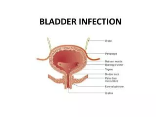
Is a hollow musculomembraneous organ that varies in form,size and position depending on the amount of urine it contains. • Small and globular,when contracted and lies on the pubic bone. • Carnivores-It extend into the abdomen • Larger animals-it is confined to the pelvic cavity. • During filling,it enlarges gradually and become pear-shaped.
The bladder can be divided into cranial vertex , Intermediate body and caudal neck which is continuous with the urethra. • The bladder is supported by double layers of peritoneum. • Most of the surface of the bladder,except the caudal part of the neck of the bladder is covered with peritoneum,which continuous as the ligaments of the bladder onto the body walls.
HISTOLOGY OF URINARY BLADDERBY: NURUL SYUHADAH BT RIBUAN The bladder has same tissue layers as renal pelvis and ureter. The layers are: -Mucosa: Transitional epithelium -Submucosa: Lamina Propria -Muscularis: Layers of smooth muscle -Serosa: Visceral peritoneum
Histology of mucosa • The inner portion of urinary bladder • Transitional epithelium will change shape from cuboidal cells to squamous cells as the bladder expands. • The epithelial layer not contain blood vessels or lymphatics. • The basement is a single layer cells. • The basement membrane separate epithelium from connective tissue. Basal layer of epithelium Superficial layer of epithelium
Histology of submucosa • The epithelium lies after connective tissue called lamina propria. • It composed of areolar connective tissue (Soft tissue found in many areas including surrounding the blood vessels and nerves and forming the layer that flexibly attaches the skin to the muscles). • It also contains blood vessels,nerves and sometimes glands. Lamina propria of the mucosa
Histology of muscularis • Underneath of submucosa of bladder is a layer called muscularis. • It consists of 3 layer of smooth muscles: inner longitudinal,middle circular and outer longitudinal. • Contraction of these muscles expels the urine from the bladder.
Histology of serosa • Serosa is covering the upper region of the bladder. • Serosa composed of simple squamous epithelium overlying a small bit of connective tissue. • The outer layer of bladder is adventitia which composed of connective tissue. • The serosa is visceral peritoneum. • Beyond of the serosa covering of bladder is perivesical fat. • This is a layer of fat surrounding bladder.
Bulbourethral Gland(anatomical aspect)BY: NUR NABILA BT MOHD ROZAINI
Also known as Cowper’s glands. • One of the accessory gland for male reproductive system. • Paired glands located on either side of the urethra. • Tubular or tubuloalveolar gland. • Surrounded by striated muscle. Horse’s Bulbourethral glands, dorsal view
The glands have ducts that empty into the urethra, the tube through which both urine and semen pass. • Composed of several lobules held together by a fibrous covering. • Lined by columnar epithelial cells, opening into a duct that joins with the ducts of other lobules to form a single excretory duct. • This ducts opens into the urethra at the base of the penis.
All domestic animals have bulbourethral glands except dog • The glands are compound tubuloalveolar glands ,structurally resemble mucus secretory glands • Simple columnar epithelium ,which varies in height depend on functional state of gland,is under control of testosterone • Smaller excretory ducts from secretory units lined with secretory cellswhile larger excretory ducts exhibit pseudostratified or stratified columnar epithelium
TUBULAR SECRETORY UNITS EXCRETORY DUCT • The glands exhibit either acinar secretory units or tubular secretory units • The secretory cells are cuboidal,low columnar or squamous ,and light staining • Fibroelastic capsule surrounds these glands contain connective tissue,smooth muscle fibers and skeletal muscle fibers in the interlobular connective tissue septum • Connective tissue septa from capsule divide the gland into several lobules CONNECTIVE TISSUE SEPTUM ACINAR TUBULAR UNITS SKELETAL MUSCLE FIBERS(L) SKELETAL MUSCLE FIBERS(T) CONNECTIVE TISSUE CAPSULE
Mouse Bulbourethral Glands • Bulbourethral glands are androgen-regulated accessory glands that are analogous to Cowper’s glands in humans • The bulbourethral glands in mice are multilobular.Each lobule composed of acini that opens into centrally located canal.Thincapsule of fibrous connective tissue surrounds and separates the acini.Theacini composed of epithelial cells.
In secretory state-epithelium has abundant ,foamy cytoplasm • In resting state—the cytoplasm is eosinophilic(appearance of cell) and finey granular. • Secretory cells are found mainly in body and resting cells are found in tail.Theductular epithelium is cuboidal. • Older mice may develop cystic bulbourethral glands.
ACINI TUBULAR GLAND lined by tall columnar epithelium SKELETAL MUSLE
BULBOCAVERNOSUS MUSCLE Histology of male genital organ in mouse
FUNCTION OF URINARY BLADDER & BULBOURETHRAL GLAND BY: AZREENASHAFIQAH BT AZMEE
URINARY BLADDER • An organ that collects urine excreted by the kidneys before disposal by urination. -Urine enters the bladder via the ureter and exits via the urethra.
BULBOURETHRAL GLAND • Male sexual glands. • Located below the prostate and discharge a component of the seminal fluid into the urethra. • Secretions provide fluid to lubricate and clean the lower urethra, which may have residue left from its use during urination.
During sexual arousal each gland produces a clear, salty, viscous secretion known as pre-ejaculate. • This fluid helps to lubricate the urethra for spermatozoa to pass through, neutralizing traces of acidic urine in the urethra,and helps flush out any residual urine or foreign matter. • It is possible for this fluid to pick up sperm, remaining in the urethral bulb from previous ejaculations, and carry them out prior to the next ejaculation. • It also produces some amount of prostate-specific antigen (PSA), and Cowper's tumors may increase PSA to a level that makes prostate cancer suspected.