Download
1 / 54
540 likes | 700 Views
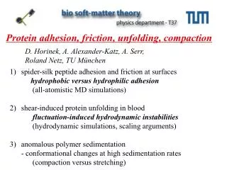
Protein adhesion, friction, unfolding, compaction. D. Horinek, A. Alexander-Katz, A. Serr, Roland Netz, TU München. spider-silk peptide adhesion and friction at surfaces hydrophobic versus hydrophilic adhesion (all-atomistic MD simulations) shear-induced protein unfolding in blood

E N D
Protein adhesion, friction, unfolding, compaction D. Horinek, A. Alexander-Katz, A. Serr, Roland Netz, TU München • spider-silk peptide adhesion and friction at surfaces hydrophobic versus hydrophilic adhesion (all-atomistic MD simulations) • shear-induced protein unfolding in blood fluctuation-induced hydrodynamic instabilities (hydrodynamic simulations, scaling arguments) 3) anomalous polymer sedimentation - conformational changes at high sedimentation rates (compaction versus stretching)
Forces at Hydrophobic Interfaces E. E. Meyer, K. J. Rosenberg, J. Israelachvili PNAS2006, 103, 15739 Hydrophobic forces act between particles whose surfaces do not posess polar groups, regardless of the exact chemical composition. Hydrophobic forces give rise to many different phenomena, Short-ranged versus long-ranged Theoretically, hydrophobic forces are not uniquely defined.
consider proteins as materials
Major ampullate silk (dragline) Flagelliform silk (capture spiral) Thomas Scheibel TUM Biochemistry Orb weaving spiders produce various silks
Thomas Scheibel TUM Biochemistry Andreas Bausch TUM Biophysics
Structural building blocks of spider silk ductile / amorphous crystalline unknown function sequence from the two dragline proteins of the garden spider A. diadematus [ ] 150 Thomas Scheibel TUM Biochemistry Single motifs are repeated up to several hundred times in spider silk proteins. Universal protein: hydrophobic/hydrophilic, unstructured and motifs
diamond-surface (Garrido/Walter/Stutzmann) H-terminated diamond OH-terminated diamond Single-molecule protein-diamond-interaction Thorsten Hugel TUM Medical Engineering AFM-tip with one spider-silk molecule NH2 NH2 NH2 NH2 NH2 PEG PEG spider silk (Scheibel) C16: MASMTGGQQMGRGSM(GSSAAAAAAAASGPGGYGPENQGPSGPGGYGPGGP)16
AFM results, hydrophobic surface (Hugel, TUM) plateau-length-distrib. plateau-force-distrib. average 58 pN • strong adsorption, yet small friction
F R FN FT if applied tangential force FT smaller than rate-dependent frictional resistance, polymer sticks; --> angle self-adjusts Serr, Netz, EPL 73, 292 (2006) Vertical pulling at constant speed, low friction Vertical pulling at constant speed, high friction
MD Simulations (Dominik Horinek) water (SPC) peptide fragment (Gromos96) H-terminated diamond surface (Gromos96) OH-terminated diamond For simulations, the spider silk C16 motif is cut in three pieces: GSSAAAAAAAASGPGGYGPENQGPSGPGGYGPGGP GSSAAAAAAA fragment 1GPGGYGPENQGPSGPGGYGPGGP GSSAAAAAAAASGPGGYGPENQGPSG fragment 2 2GPGSSAAAAAAAASGPGGYGPENQGPSGPGGYGPGGP fragment 3 Alkane SAM
a Simulations of AFM Desorption of Spider Silk from Surfaces b AFM tip pulled group solid peptid is pulled from surface via a moving spring attached to the terminal group high mobility on surface!
from lateral diffusion of adsorbed peptides Single-molecule-friction on Hydrophobic Diamond friction coefficient per length ≈ 0.05 kg/sm friction force for 100nm peptide at v=1 m/s : 0.05 kg/sm 1 m/s 100nm = 5 fN !!! Too small for the AFM ! 3 center-of-mass trajectories of spider silk at different tip elevations. hydrophobic binding is self-lubricating
Desorption Forces from MD at various pulling ratesHorinek, RRN, PNAS (2008) a Fdes = k (zspring -zAA) b 10 pulling rate 10 m/s c 1 pulling rate 1 m/s d 0.1 pulling rate 0.1 m/s hydrophobic surface average plateau force: 54 pN (experimental: 58 pN with NaCl)
spontaneous desorption vertical pull U is a result of partial compensation of large individual energies first 3 contributions nearly compensate energy decomposition - hydrophobic attraction forget simple-minded theories concentrating on one aspect !!
hydrophilic OH-terminated diamond Pulling rate 0.1 m/s Large hysteresis ! desorption (at most) doubled on hydrophilic surface large friction due to breaking and reformation of hydrogen bonds !!
spider silk friction for lateral pullingAndreas Serr • Hydrophobic diamond • pulling rate 8 m/s
Spider silk friction • Hydrophilic diamond (50% OH) • pulling rate 1 m/s
experimental friction forces: simulation 0.1m/s -> 5pN simulation 0.1m/s -> 200pN Single-molecule peptide friction bulk water (perfect match with exp.) • mobility per monomer hydrophobic diam hydrophilic diam 30-fold friction increase for hydrophilic surface: driven diffusion in corrugated binding potential of 6kBT (Frenkel-Kontorova-Tomlinson) o.k. agreement with exp. data peptide glides on vacuum: „hydrophobic binding is self-lubricating“
- adhesive proteins bind to BOTH hydrophilic and hydrophobic • surfaces strongly (5 kBT per amino acid) • nano-friction on hydrophobic/philic substrates is very different • (effective adhesive properties depend on binding free energy • AND surface friction ! gecko, scotch tape) • in all cases, effective interaction involves direct • interactions as well as water-ordering effects!
hydrophobic / philic homopeptides Salt Effects Hugel lab F N Specific Ion Adsorption at Hydrophobic Solid Surfaces D. Horinek / RRN, PRL99, 226104 (2007)
pressure between 2 hydrophobic surfaces from Poisson-Boltzmann:screenable contribution to hydrophobic attraction 1 mM salt: weak but long-ranged 100 mM salt: strong but short-ranged DOES NOT YET EXPLAIN PEPTIDE ION SPECIFICITY!
blood functions: - oxygene - transport (& Hemoglobin) - nutrient - transport (glucose, amino-acids, fat ....) - waste - transport (CO2l urea, lactatic acid ...) - immuno reactione ( lymphocytes, antibodies ...) - signal - transduction (hormons ...) - regulation of temperature and pH of body - coagulation, vascular repair
capillaries connect arteries and veins they are 5-10 microns thick and are lined by a single-cell-layer: the endothelium
action since the endothelial layer is thin, it ruptures easily ! the von-Willebrand-factor (vWF) helps fixing capillaries
Blood von-Willebrand Faktor (fibers !!!) Transport Docking Fusion Intracellular Vesicels (packaged proteins) von-Willebrand Faktor (globular !!!) vWf unfolds in shear
von-Willebrand desease caused by unspecific deficiency of vW-factor bleeding of small vessels with shear rates > 1000 s-1
monomer (2500 aminoacids) dimer multimer (a few hundred units) the vWf is the largest watersoluble protein in the body --- why ???
Large globular structure ~ 25x6.5 nm Rod + central nodule ~ 30 + 6 nm von-Willebrand factor (vWf) Lines 120 nm apart Fowler et al vWf bietet Bindungsstellen für Kollagen und Blutplättchen, Kollagen schaut aus kaputten Blutgefäßen heraus!
Was stimuliert die Entfaltung des vWf ?? Hypothese: Scherfluss in kleinen Blutgefäßen bewirkt Entfaltung des Proteins! R Hagen-Poiseuille Gesetz für Strömung im Rohr: Flüssigkeitsstrom geht wie R4 Strömungsgeschwindigkeit ist Null an der Wand Scherung verformt Proteine und Blutkörperchen Experimentelle Untersuchung an künstlichen Blutgefäßen!
Flow-chamber Chip - Wixforth&Schneider, Augsburg 40mm 200µm 1mm Surface Acoustic Wave (Nanopump) Hydrophobic Surface High Frequency Input (Source of SAW) V = 8µl LiNbO3 (Piezoelectric) Hydrophilic Channel
unfolding occurs also in bulk (without collagen substrate) Schneider/Wixforth (Augsburg) relaxation into globular state once shear is turned off
Quantitative experimental measurements linear vWf extension vWf adhesion efficiency on collagen substrate vWf unfolds abruptly at shear rates of about 3000 s-1 (close to shear rates in capillaries) adsorption on collagen starts at about the same shear rate! Seek deeper understanding through theoretic modeling !
length and time scales (microns and milliseconds) require coarse-grained simulations techniques! coarse-grained description - few effective interactions - only implicit solvent atomistic resolution - detailed force fields - including explicit water
Hydrodynamics at low Reynolds numbers Stationary Navier-Stokes equation , , If the Reynolds number human bacteriumsinking cylinder one obtains the creeping flow equation. H2O: = 0.001 Pa s; = 1000 kg/m3 v = 1 m/s l = 1 m v = 10-5 m/s l = 1 Re = 10-5 v ~ 10-7 m/s l = 1 Re = 10-7 Re = 106
flow-field due to point-force at origin: (Oseen-Tensor) for many particles the superposition principle is valid: invert to get forces for prescribed solvent velocity distribution !! Next: add thermal noise
Hydrodynamic Brownian simulation techniques Velocity of i-th particle: deterministic force Random force Mobility matrix: self mobility: hydrodyn. interact. equivalent to Smoluchowski equation for particle distribut. W(rj,t) : with solution:
simple model for protein coil-globule transition Alfredo Alexander-Katz, RRN attractive Lennard-Jones potential between all monomers
globule in shear flow, =2.5, =1.2 Alfredo Alexander-Katz, RRN
unfolding dynamics Rg2 ~ * time (a. u.) shear-induced unfolding unfolding becomes abrupt for strongly folded proteins (in agreement with experiments)
protrusion-instability mechanism is fundamentally different from classical droplet instability (Taylor 1934) out(outside viscosity) in - critical shear rate is temperature dependent - Taylor: stable for in / out > 4 - instability occurs on small length scales - final results depends on lower spatial cutoff
minimal model for shear-induced globule unfolding: “force balance on protrusions” cohesive force on protrusion (sharp interface, diffuse interface) from equipartition theorem lf=kBT --> „typical“ protrusion length relative velocity sphere/solvent # monomers shear-force on protrusion -free draining (with slip) friction coefficient of one monomer -hydrodynamic case (no slip) --> typical cohesive force on protrusion fcoh
critical protrusion length fcoh = fshear free draining hydrodynamic
scaling of critical shear rate (with hydrodynamics) : L: protein contour length a: protein monomer radius to unfold a protein with typical cohesion energy in a capillary vessel one needs huge monomers with a radius of 10 nm, close to vWf enormously large monomer size !!! =2kBT, N=100, =1000s-1, ----> a = 10nm !! now connect to classical hydrodynamic instability theory (Taylor, Kelvin-Helmholtz) and assume protrusions are controlled by surface tension /a2 and instability at small length scales !! A. Alexander-Katz, RRN: PRL (2006), PNAS (2007) ……..
G: sedimentation force per monomer N: monomer number polymer separation in the ultracentrifuge: sedimentation anomaly at large driving fields • velocity v = GN • mobility R N • velocity v ≈ G N1- sedimentation rate S = v/G ≈ N1-
why gel-electrophoresis is used for separating DNA (and not the ultracentrifuge) - sedimentation rate of polymers goes down at high rotor speeds - crossover is polymer-length dependent! EJ Ralston/VN Schumaker 1974 / 1979 circular episome 1338 DNA Sedimentation rate linear episome 1338 DNA at low concentrations Rotor speed
theoretical explanation by Zimm (1974): • free ends of polymer are typically peripheral • receive more drag in sedimenting flow • stretched arch shape is produced • sedimentation coefficient goes down • NULL EFFECT PREDICTED FOR CIRCULAR CHAINS • within pre-averaging approximation • - story ended in 1979 flow
Crumpling and stretching of sedimenting flexible chain (Schlagberger, Netz, PRL 2007) motion