Download
1 / 24
240 likes | 652 Views
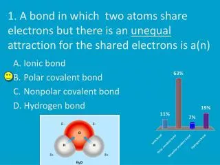
The name is bond---H bond Akhil Khanal February 2006. Early evidence of hydrogen bonding. Many elements form hydrides Plot BP of hydrides of Group 4 elements BP increases as we go down a gp Van der Waals forces Plot BP of hydrides of gp 5,6, and 7 Similar results

E N D
Early evidence of hydrogen bonding • Many elements form hydrides • Plot BP of hydrides of Group 4 elements • BP increases as we go down a gp • Van der Waals forces • Plot BP of hydrides of gp 5,6, and 7 • Similar results • EXCEPT the hydride of the first element • in each of groups 5,6, and 7 • NH3, H2O, and HF must be having some • additional intermolecular forces of attraction
Hydrogen attached directly to most electronegative atoms • Partial positive charge of hydrogen • Partial negative charge on the electronegative atoms • as well as at least one lone pair • The partially positive hydrogen strongly • attracted to the lone pair • Interaction much stronger than • dipole-dipole interaction • Ethanol (BP = 78.5 C) • Methoxymethane (BP = -24.8 C) • 100-fold elevation of BP
Hydrogen bonding in water • H-bond partly electrostatic (90%), partly covalent (10%) • Approximated by the following bonds: • a (A-H B); b (A- H+---B: ionic);c(A- H----B+: covalent);d(A+ H----B: ionic);e (H- A---B+: covalent) • = a +b +c +d +e • X-Ray spectroscopic evidence suggests that these interactions shift within a femtosecond • Nuclear quantum effects strengthen the H-bond by increasing the dipole moment • Donor hydrogen stretches away from its oxygen • Acceptor lone-pair stretches away from it oxygen • Both oxygens being pulled towards each other Cute Movie: http://www.northland.cc.mn.us/biology/Biology1111/animations/hydrogenbonds.html
H-bonds direction, lengths, angles, and strength • H-bond possesses direction and is generally asymmetric • Bond strength depends on angle and length • Small changes from linearity (20º) have minor effect in strength • H-bond strength, on the other hand,decreases exponentially with distance • There is a tradeoff between H-bond and covalent bond strength • stronger the H····O bond, the weaker the O-H covalent bond, • and shorter the O····O distance • Weakening of the covalent bond is a good indicator of strengthening of H-bond energy • Cooperativity and anti-cooperativity in H-bonding
How would you calculate the strength of an H-bond? • Between 3 and 9 kcal/mol • Dissociation rate constants of 4 X 10¹⁰ to 2 X 10⁶ s-1 from transition state theory R-O-H-----ORH ROH + ROH [R-O-H-----ORH] Keq = [ROH]2/ If Keq=1, G=0; if Keq= 10, G=1.3 kcal/mol, if Keq= 100, then G= 2.6 kcal/mol • But it is more complicated than this for biological systems in water
H-bond Inventory • Hard to determinethe exact strength because of H-bonding to water • R-O-H----ORH ROH + ROH • Atypical H-bonding reaction • E-XH---OH2 + OH2---B-S E-XH---B-S +H2O---H2O • Count the number of H-bonds on each side • H-bond isoenthalpic • Remember entropic advantage!
Complications • A + B A.B • A has 3 rotational and 3 translational Degree of Freedom (DoF) • B has 3 translational and 3 rotational DoF • A.B only has 3 translational and 3 rotational DoF • Net loss of 3 degrees of freedom • For small molecules, 3 DoF equal 12-16kcal/mol • Therefore, H-bonds are worth more than 3-9 kcal/mol because they have to • overcome their 12-16 kcal/mol of freedom loss
Different types of H-bonds • Common elements that form H-bonds • S, O, N, F • CH H-bond RHO----RCOH+COR’ • -cation interaction
Low Barrier Hydrogen bonds (LBHB) • H-bond strength depends on its length, linearity, microenvironment, and the pKa values of the the H sharing components • H-bonds in water are relatively weak because of pKa mismatch between H3O+ (-1.7) and H2O (15.7) • The proton in the structure is tightly associated with the OH- as a water molecule • In gas phase, dielectric constant is low • Hydrogen bonds between heteroatoms with matched pKa values can be 2.5 Å and very strong (25-30 kcal/mol)
Detection of LBHB • Low field proton NMR signal • O-H and N-H bond length gets longer, H gets deshielded • Chemical shifts are 18-22 ppm in LBHB • Eg., proton NMR spectra of monoanion of cyclopropane-1,1-dicarboxylic acid • Deuterium fractionation factor of the central hydrogen • O-H and N-H bond length increases, bond order decreases • Discrimination against deuterium increases 0.3-0.5 Enz-H + Dsolvent Enz-D +Hsolvent = [Enz-D][Hsolvent]/[Enz-H][Dsolvent]
LBHB in enzymes • Change the substrate pKa to match that of the amino acid • H-bond between lactate (15) and His-195 (6) • Mismatch of 9 pH units • During the rxn, pKa of lactate crosses that of histidine • LBHB forms and activation energy of the reaction is lowered • Elimination of mismatch of 9 pH units = 4.5 orders of magnitude • acceleration of rate
Other examples • Ketosteroid Isomerase • Triose-phosphate Isomerase • Citrate Synthase • Mandelate Racemase • Cleland et al., JBC, 1998,(273), 25529-25532 • chymotrypsin
H-bonds in Proteins I. H-bonds contributing to structure and folding II. H-bonds contributing to catalysis
-helix • i+4, i+3, i+5 • Dipole moment • + charge at N-terminii, - charge at COOH • Negative molecules (PO42- groups) bind at N-terminii
-sheets • Antiparallel -sheets • 10-14 atoms in a ring • H-bonds linear • Stability? • Parallel -sheets • 12 atoms in a ring • H-bonds not 180 • Stability?
Peptide H-bonds • Strength of H-bond = 3-9 kcal/mol • Is this the energy that it provides towards protein folding? • Remember, folded protein loses entropy • Entropic cost of freezing one aa residue is 1.2 kcal/mol • What then drives protein folding? H-bonds? Hydrophobics? • Hydrogen bond inventory • about 0 ± 1 kcal/mol for peptide bond in water • A protein H-bond network is complex • Protein forms 75% of it H-bonds • In a 100 aa protein this represents 70 kcal/mol of energy towards folding • Note: folded proteins are generally 5-10 kcal/mol more stable than unfolded ones
Thermolysin-inhibitor complexes • Crystal structures of thermolysin • first with inhibitor A (has NH) • second with inhibitor B (has O) • Differ by only one H-bond • Contribution of that one additional H-bond?
H-bonds in catalysis • Engineered enzyme relative to the WT has essentially the same KM but kcat decreases • 3000-fold!
Ligand binding H-Bonds Hydrogen bond types Steiner, T. Angew. Chem. Int. Ed. 2002, 41, 48.
Few papers you all might find enjoyable (or maybe not…) • Martin et al., Nature Structural biology, 1999, (6), 403-406. • Robert Baldwin, Journal of Biological Chemistry, (2003), (278), 17581-17588. • Cleland et al., Journal of Biological Chemistry, 1998, (273), 25529-25532. • Fleming et al., Protein Science, 2006, (14), 1911-1917.