Vision: How do we see?
Vision: How do we see?. Or, how does radiation get converted for interpretation by our brain?. Electromagnetic Radiation. “Visible light” is a small part of this spectrum!. Why do animals “see”?. Why not just smell? Or hear? Or feel? Or taste? What is an advantage of vision over others?

Vision: How do we see?
E N D
Presentation Transcript
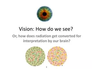
Vision: How do we see? Or, how does radiation get converted for interpretation by our brain?
Electromagnetic Radiation • “Visible light” is a small part of this spectrum!
Why do animals “see”? • Why not just smell? Or hear? Or feel? Or taste? • What is an advantage of vision over others? • But how did it happen? www.youtube.com/watch?v=lEKyqIJkuDQ
How do we see? • Vertebrates (and other animals) have receptors that are stimulated by light • The eye focuses light on these receptors • Millions of these receptors allow an “image” of the environment to be formed • What does this “image” look like? Depends on what stimulates your receptors!
Structure of the Vertebrate Eye • You will (or did) see these in lab Fig. 17.17
Structure of the Vertebrate Eye • Outer layer = ________ • Thick connective tissue • Attachment site for extrinsic eye muscles • Anterior portion is clear = _______ • Middle layer = _____with 3 regions • Choroid: pigmented, vascularized • Ciliary body: • Ciliary muscle – smooth muscle attached by suspensory ligament to lens • Ciliary processes – produce aqueous humor • Iris: smooth muscle that controls size of ____
Structure of the Vertebrate Eye • Inner layer = retina with 3 cell layers • Photoreceptor cells (rods, cones) • Horizontal cells and bipolar cells • Synapse with rods and cones • Horizontal cells inhibit photoreceptors, adjust output • Amacrine cellsand ganglion cells • Amacrine cells adjust output of bipolar cells • Axons of ganglion cells run along surface of retina, joining to form optic nerve (at optic disk)
Structure of the Vertebrate Eye Note positioning Fig. 17.17
Structure of the Vertebrate Eye • Inner layer = retina with 3 cell layers • In some vertebrates, retina is indented, forming a fovea • Fovea = point at back of eyeball where light converges –
How do we get light to focus on the back of the eye? • Depends on where you live: • Terrestrial: cornea refracts light, lens refines • Aquatic: lens refracts light, cornea does little • Why? Fig. 17.20
What about things at different distances away? • Accommodation– • Can do so with different means Iris muscles change lens shape Corneal muscles pull cornea against lens Muscles change lens position Ciliary muscles change lens shape Fig. 17.21
Photoreceptor Cells • Rods – very sensitive to low light levels • “Night vision” • Cones – different types sensitive to different wavelengths of light Light-absorptive pigments here! Rhodopsin in rods Photopsins in cones
Photoreceptor Cells • Many rods converge on one bipolar cell • Nocturnal species • Cones have one-to-one synapse with bipolar cell Fig. 17.23
Photoreceptors PDE = phosphodiesterase GMP = guanosine monophosphate Stimulated photoreceptor cell decreases release of neurotransmitter (= glutamate) to bipolar cell - what is happening in the dark?
Cones and Color Vision • 4 cone cells (tetrachromatic) – based on opsin • UV/Violet(370 nm) – Blue (445 nm) – Green (508 nm) – Orange/Red(650 nm) • Many fishes, turtles, lizards, birds • Oil droplets filtering short λ • Brain assigns colors human vision (R+G+B) only UV vision (UV) “bee vision” (UV+G+B) “bird vision” ( UV+R+G+B)
Cones and Color Vision • Amphibians with color vision are trichromatic • Some have lost color vision
Cones and Color Vision • Mammals – Dichromatic (≠ B&W) • Violet/Blue – yellow/ orangecone cells • Color-blindness in green, orange, red regions • Nocturnal ancestry? • Exceptions are humans and apes - secondarily derived trichromatic vision • No oil droplets
After-images • What happened?
After-images • First, why does the screen look “white? • Photoreceptors get “bleached”, and our brain …
Depth Perception • Trade-off between breadth of visual field and depth perception • Monocular vision – • Binocular vision – Fig. 17.24
Depth Perception • Binocular vision: half of info goes to same side of brain, half goes to other via … • Comparisons made in … Fig. 17.25
Integrating Visual Information Fig. 17.26
Infrared Perception • IR = narrow range of wavelengths of electromagnetic radiation, NOT heat • IR receptor (thermoreceptor): SSO • Vampire bats, boas, pit vipers • Free nerve ending in skin • Absorbs IR radiation → Warmed → Info to brain • Only needs ± 0.003°C • Pit vipers can detect mice 30 cm away http://www.youtube.com/watch?v=lySW2-eYilg