Download
1 / 38
380 likes | 412 Views
Learn about the divisions of the Central Nervous System, including the Brain, Brain Stem, Limbic System, and Cerebral Cortex. Explore functions and key areas like the Thalamus, Hypothalamus, and more.

E N D
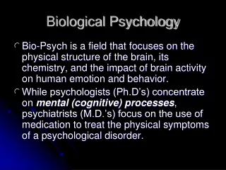
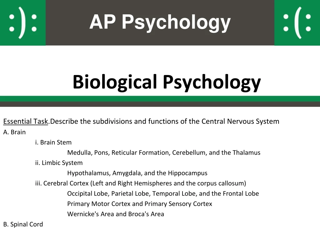
AP Psychology Biological Psychology Essential Task.Describe the subdivisions and functions of the Central Nervous System A. Brain i. Brain Stem Medulla, Pons, Reticular Formation, Cerebellum, and the Thalamus ii. Limbic System Hypothalamus, Amygdala, and the Hippocampus iii. Cerebral Cortex (Left and Right Hemispheres and the corpus callosum) Occipital Lobe, Parietal Lobe, Temporal Lobe, and the Frontal Lobe Primary Motor Cortex and Primary Sensory Cortex Wernicke's Area and Broca's Area B. Spinal Cord
Endocrine System Evolutionary We are here Building Blocks Genetics Biological Psychology Neurons Neurotransmitters Nervous System Central Nervous System Peripheral Nervous System Motor Sensory Spinal Cord Brain Autonomic Somatic Brain Imaging Sympathetic Parasympathetic
Essential Task: CNS Outline A. Brain i. Brain Stem Medulla, Pons, Reticular Formation, Cerebellum, and the Thalamus ii. Limbic System Hypothalamus, Amygdala, and the Hippocampus iii. Cerebral Cortex (Left and Right Hemispheres and the corpus callosum) Occipital Lobe, Parietal Lobe, Temporal Lobe, and the Frontal Lobe Primary Motor Cortex and Primary Sensory Cortex Wernicke's Area and Broca's Area B. Spinal Cord
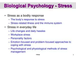
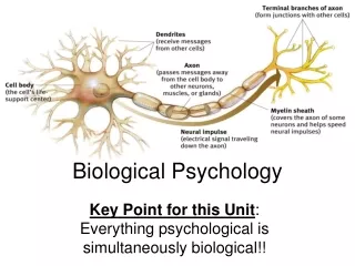
The Somatic Nervous System • Consists of neurons that communicate between the body and the brain • Motor Neurons • Neurons that carry messages from the spinal cord or brain to muscles and glands
The Autonomic Nervous System • Sympathetic division • Most active when you are angry, afraid, or aroused • Increases heart rate and breathing • Stops digestion • “Fight-or-flight”
The Autonomic Nervous System • Parasympathetic division • Calms body • Produces effects opposite to those of the sympathetic division • Reduces heart rate and breathing • Restores digestion • “Rest and Digest”
The Hypothalamus Has Central Control of the ANS • When someone experiences a stressful event, the amygdala, an area of the brain that contributes to emotional processing, sends a distress signal to the hypothalamus. This area of the brain functions like a command center, communicating with the rest of the body through the nervous system so that the person has the energy to fight or flee. • The hypothalamus is involved in the coordination of ANS responses • One section of the hypothalamus seems to control many of the "fight or flight" responses; another section favors "rest and digest" activities
Language Aphasiais an impairment of language, usually caused by left hemisphere damage either to Broca’s area (impaired speaking) or to Wernicke’s area (impaired understanding).
Specialization & Integration Brain activity when hearing, seeing, and speaking words
Association Areas More intelligent animals have increased “uncommitted” or association areas of the cortex.
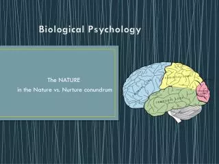
Our Divided Brain Our brain is divided into two hemispheres. The left hemisphere processes reading, writing, speaking, mathematics, and comprehension skills. In the 1960s, it was termed as the dominant brain.
Hemispheric Specialization • Corpus Callosum • Fibers that connect the two hemispheres • Allow close communication between left and right hemisphere • Each hemisphere appears to specialize in certain functions
Hemispheric Specialization People with intact brains also show left-right hemispheric differences in mental abilities. A number of brain scan studies show normal individuals engage their right brain when completing a perceptual task and their left brain when carrying out a linguistic task.
Splitting the Brain A procedure in which the two hemispheres of the brain are isolated by cutting the connecting fibers (mainly those of the corpus callosum) between them. Corpus Callosum
Split Brain Patients With the corpus callosum severed, objects (apple) presented in the right visual field can be named. Objects (pencil) in the left visual field cannot.
Fun with Your Hemispheres • Rotate your dominant hand in one direction while at the same time rotating the opposite foot in the other direction. • No problem since controlled by two hemispheres • Now, rotate your dominant hand in one direction while at the same time rotating the foot on the same side in the other direction.
Older Brain Structures The Brainstemisthe oldest part of the brain, beginning where the spinal cord swells and enters the skull. It is responsible for automatic survival functions.
Brain Stem The Medulla [muh-DUL-uh] is the base of the brainstem It controls autonomic functions and relays nerve signals between the brain and spinal cord. • respiration • blood pressure • heart rate • reflex arcs • vomiting
Brain Stem Pons and inside that the (Reticular Formation)isa nerve network in the brainstem that plays an important role in controlling arousal. • It is involved in motor control and sensory analysis... for example, information from the ear first enters the brain in the pons. It has parts that are important for the level of consciousness and for sleep. The Reticular Formation controls: • Attention • Cardiac Reflexes • Motor Functions • Regulates Awareness • Relays Nerve Signals to the Cerebral Cortex • Sleep
Brain Stem The Medulla [muh-DUL-uh] is the base of the brainstem that controls heartbeat and breathing. Reticular Formationisa nerve network within the brainstem that plays an important role in controlling arousal.
Brain Stem The Thalamus [THAL-uh-muss] is the brain’s sensory switchboard, located on top of the brainstem. It directs messages to the sensory areas in the cortex and transmits replies to the cerebellum and medulla.
Cerebellum The “little brain” attached to the rear of the brainstem. It helps coordinate voluntary movements and balance.
The Limbic System The Limbic Systemisa doughnut-shaped system of neural structures at the border of the brainstem and cerebrum, associated with emotions such as fear, aggression and drives for food and sex. It includes the hippocampus, amygdala, and hypothalamus.
Amygdala The Amygdala [ah-MIG-dah-la] consists of two almond-shaped neural clusters linked to the emotions of fear and anger.
Hypothalamus The Hypothalamus lies below (hypo) the thalamus. It directs several maintenance activities like eating, drinking, body temperature, and control of emotions. It helps govern the endocrine system via the pituitary gland.
Reward Center Rats cross an electrified grid for self-stimulation when electrodes are placed in the reward (hypothalamus) center (top picture). When the limbic system is manipulated, a rat will navigate fields or climb up a tree (bottom picture). Sanjiv Talwar, SUNY Downstate
The Cerebral Cortex The intricate fabric of interconnected neural cells that covers the cerebral hemispheres. It is the body’s ultimate control and information processing center.
Structure of the Cortex Each brain hemisphere is divided into four lobes that are separated by prominent fissures. These lobes are the frontal lobe (forehead), parietal lobe (top to rear head), occipital lobe (back head) and temporal lobe (side of head).
Functions of the Cortex The Motor Cortex is the area at the rear of the frontal lobes that control voluntary movements. The Sensory Cortex (parietal cortex) receives information from skin surface and sense organs.
Visual Function The functional MRI scan shows the visual cortex is active as the subject looks at faces.
Auditory Function The functional MRI scan shows the auditory cortex is active in patients who hallucinate.
The Spinal Cord • Complex cable of nerves that connects brain to rest of the body • Carries motor impulses from the brain to internal organs and muscles • Carries sensory information from extremities and internal organs to the brain • 400,000 people a year in US suffer either partial or complete paralysis
The Spinal Cord • The spinal cord controls some protective reflex movements without any input from the brain