Download
1 / 30
300 likes | 851 Views
Sensory and Neurological Disorders. Dr. Kline FSU-PC. I. Sensory Disorders. Are comprised of deficits in sensory modalities resulting from neurological damage to the CNS. A. Visual disorders.

E N D
Sensory and Neurological Disorders Dr. Kline FSU-PC
I. Sensory Disorders • Are comprised of deficits in sensory modalities resulting from neurological damage to the CNS.
A. Visual disorders • 1. Scotomas- small blind spots in the visual field resulting from damage to the primary visual cortex . • **May also occur temporarily during migraines. • Most people are unaware of these because of nystagmus, constant tiny involuntary eye movements that “fills in the missing information.” • Blind spots become obvious, if eyes are held still.
2. Cortical blindness • Patients with complete damage to the primary visual cortex (V1) report being totally blind. • Despite reporting being blind, these patients can grab a a moving object or track a moving light. Patients report being unaware of their ability to do this. • The ability of cortically blind people to perform visually mediated tasks without conscious awareness is called blindsight.
Cortical Blindness case study: D.B. • D.B, was blind in his left visual field (LVF) because his right occipital lobe had been surgically removed. He could see images in his right visual field (RVF) because his left occipital lobe was intact. • “Even though the patient had no awareness of “seeing” in his blind field, evidence was obtained that he could reach for visual stimuli in his left field with considerable accuracy, could differentiate the orientation of a vertical line from a horizontal or diagonal line, and could differentiate the letters X and O.” • D.B. showed great surprise at being told he was accurate at these tasks.
3. Visual Agnosias: • Refers to inability to recognize objects, their pictorial representations, or to draw or to copy them. • These people are not blind, they can point to objects & describe their features. However, they can’t determine what the object is.
Example of patient with visual agnosia • The patient was shown a key. He could describe the individual components of the key, but could not say what the item was. • When shown a stethoscope, he said “a stethoscope is a long tube with a round thing at the end.” • When told it was a stethoscope he would agree with the doctor, but could not recognize the object himself. When told it might not be a stethoscope his response was that if the doctor didn’t think it a stethoscope he would not either because he lacked any confidence in his ability to recognize and name the object.
Types of visual agnosias • a. Visual Object Agnosia: the patient can see the object, but is unable to name it, demonstrate its use, or remember having seen it before. • E.g., One patient described a bicycle as “a pole with two wheels, one in front and one in back.” • Lesion is supposed to be in left occipital lobe in secondary cortex, although it is most common for the damage to be bilateral.
b. Visual Agnosia for drawings: • Effects recognition of a variety of drawn stimuli, including realistic representations of simple objects, geometric figures, meaningless forms, incomplete figures, & abstract drawings. • The lesion producing this condition is in secondary visual cortex.
c. Prosopagnosia • First noticed in 1947 when three patients with head trauma described the inability to recognize faces although they were able to recognize objects, forms, & colors. • People with prosopagnosia cannot identify faces (& some complex objects). They often recognize others by their voice or gait. • Damage in prosopagnoisa occurs in two types of cases: • **bilateral damage to the inferior temporal lobe** • **unilateral damage to right posterior parietal lobe**
4. Motion Blindness • Patients with this disorder can see objects, but have trouble determining whether an object is moving or stationary. • **For these people life is a series of snap shots or photos. You can think of it as a series of “freeze frames.” • **The middle temporal lobe (V5) has cells that respond to movement. This is the area of damage in patients with motion blindness.
B. Somatoperceptual Disorders • 1. Astereognosia: the inability to recognize objects from touch (even if able to do so previously). Damage is to postcentral gyrus (primary somatosensory cortex). • 2. Blind touch: patients can identify the location of a visual stimulus even though they deny “seeing” it. 3. Asomatognosia:Is the loss of knowledge or sense of one’s own body & bodily condition. The person neglects part of his or her body.
Asomatognosia—lesion in postcentral gyrus. • These may be for one or both sides of the body. They do appear to be most common for the left side of the body resulting from right hemispheric lesions. Next slide—description of one of Oliver Sacks’ patients with asomatognosia.
The patient had felt fine all day and fallen asleep towards evening. When he woke up he felt fine until he moved in the bed. • Then he found, as he put it, ‘someone’s leg’ in the bed—a severed human leg!! Stunned & then disgusted, he thought one of the nurses was playing a joke on him (put a dismembered body part in bed with him). …When he threw it out of bed, he somehow came after it—and now it was attached to him. “Look at it!” he cried. “Have you ever seen such a creepy, horrible thing?… The nurse asked him to remain calm. He became irritated arguing, “Why!” The doctor then came and answered “Don’t you know your own leg?” The patient responded, “Ah, Doc!, you’re folling me! You’re in cahoots with that nurse.” Sacks responded, “Listen, I don’t think you’re well. Please allow us to return you to bed. But I want to ask you one final question. If this—this thing—is not your left leg… then where is your own left leg?” The patient, looked pale and said, “I don’t know, I have no idea, its disappeared, gone forever.”
4. Contralateral Neglect • Usually caused by right posterior parietal lobe damage, this disorder was first described in 1874. • Famous case study highlighted disorder: • Mr. P, 67 at the time of his right parietal lobe stroke, had unusual symptoms post-stroke.
Mr. P’s Symptoms: • 1. He neglected the left side of his body & world. • E.g., when asked to life the arms up, he would fail to lift the left arm. • 2. He would draw a clock face, with all the numbers crowded on the right side of the clock. 3. He ignored tactile sensations on the left side of the body. e.g., Didn’t brush hair of left side or teeth in left side of mouth.
Contralateral neglect symptoms • Global deficit--neglect of visual, auditory, & somaesthetic (somatosensory) stimulation on the side of the body and/or space opposite to the lesion. • Unilateral spatial neglect (usually left side) • Visual spatial neglect (deficit in comprehending visual space) • Dressing apraxia—dress half of the body • Paralexia-read half of a word • Paragraphia-writes only half of a word • Hemi-inattention—ignore opposite side of body • Hemi-akinesia-poverty of movement of one side of body. • Anosognosia-denial of illness or symptoms
5. Apraxia • Is a loss of skilled movement that is not caused by weakness; an inability to move; abnormal tone or posture, intellectual deterioration, poor comprehension or other disorders of movement such as tremor. • Two types: • 1. Ideomotor apraxia– patients are unable to copy movements or to make gestures (waving hello). • Damage appears to be in left posterior parietal area.
2. Constructional apraxia • Refers to a visuomotor disorder in which patients are unable to perform activities such as assembling, building, or drawing. • May result from injury to either parietal lobe; most often found in the posterior parietal region. • E.g., these patients cannot put together a puzzle.
C. Auditory Perceptual disorders • Deficit in perception of brief temporal sequences of sounds (need more time between sounds). ---Patients have difficulty with rapid sound sequences. • 2. Deficits in perceiving rapid speech (related to #1). • 3. Auditory sequencing for verbal information may be impaired. Damage is usually in the left hemispheric lesion. • 4. Cats with unilateral or bilateral lesions of the auditory cortex lack the ability to localize sounds.
Audioperceptual disorders (contd.) • 5. Auditory agnosias-impaired capacity to recognize nonverbal sounds (very few cases reported). • 6. Amusia-disruption in recognition of music (tones, melodies, or rhythms).
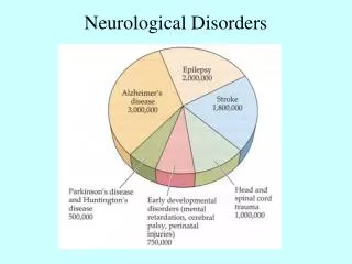
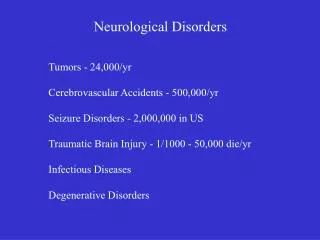
II. Neurological Disorders • The normal functioning of the CNS can be affected by a number of disorders, the most common of which are headaches, tumors, vascular problems, infections, epilepsy, head trauma, demyelinating diseases, and metabolic & nutritional diseases.
A. Vascular Diseases • Vascular diseases in the brain can produce serious—even total reduction in the flow of oxygen & glucose, resulting in critical interference with cellular metabolism. • Interference lasting longer than 10 min., results in all cells in that region dying. • These are among the most common causes of death & chronic disability in the Western world.
1. Stroke (Cerebral vascular accident) • Symptoms accompany severe interruption of blood flow to the brain. • Stroke produces an infarct (area of dead or dying tissue resulting from obstruction of blood vessels normally supplying area). • Nature of deficits depend on area of obstruction, size of blood vessels (better prognosis for small vessels than large, relative health of surrounding vessels, etc.
2. Cerebral Ischemia- insufficient supply of blood to brain, are like mini-strokes. • Decreases in blood flow result of 3 causes: • A. Thrombosis-a plug or clot in a blood vessel that remains at its point of formation. • B. Embolism -moving (clot, bubble of air, sack of cells, or fat deposit) from larger vessel into a smaller vessel. • C. Cerebral arteriosclerosis-thickening & hardening of arteries.
3. Cerebral Hemorrahage • Massive bleeding in the brain. Onset is abrupt & may be quickly fatal. • Causes: • Hypertension • Congenital defects of cerebral arteries • Leukemia • Toxic chemicals
4. Aneurysms • Vascular dilations resulting from localized defects in the elasticity of the vessel. • Most common symptom is severe headache, often present for many months to years.
B. Open-Head Injuries: • Puncture or penetration of the skull through projectiles (gunshots/missile wounds) or other moving objects. • Most people with open-head injuries do not lose consciousness & produce distinctive symptoms that may undergo rapid & spontaneous recovery. • Deficits are specialized & often resemble those of surgical excisions.
C. Closed-Head Injuries • Caused by a blow to the head (car accident, blunt instrument swung at head). • Damage at site of blow is called a coup. • With severe blow, the brain may shift & hit the opposite side of the skull producing an additional bruise (contusion) known as a countercoup.
Closed-Head Injuries (Contd.) • Finally, the brain may suffer additional damage, from the shearing of nerve fibers resulting in microscopic lesions. • Frontal & temporal areas most likely to be damaged in closed-head injuries. • These injuries are common accompanied by loss of consciousness (from damage to brainstem fibers), edema (swelling), and hemorrhaging. • Length of coma often is positively correlated with severity of damage.