Download
1 / 70
780 likes | 1.17k Views
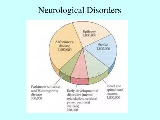
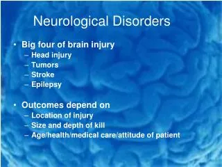
Neurological Disorders. Structural Organization. Cerebral hemispheres. Brainstem & Cerebellum. Spinal and Cranial. Spinal Cord. Nervous System Function. Brain : Central Processing Unit. Sensory Inputs. afferent. efferent. Secretion Movement. anterolateral. dorsal.

E N D
Structural Organization Cerebral hemispheres Brainstem & Cerebellum Spinal and Cranial Spinal Cord
Nervous System Function Brain: Central Processing Unit Sensory Inputs afferent efferent Secretion Movement
anterolateral dorsal Sensory Tracts
Dorsal column ipsilateral until medulla, then crosses sensation is well localized touch, vibration, pressure, Major Sensory Tracts • Anterolateral (Spinothalamic) • crosses immediately in the cord • sensation is poorly localized • itch, pain, temp
Medial Tracts some tracts cross at medulla, some don’t innervates axial muscles balance, gross motor Major Motor Tracts • Lateral Corticospinal • crosses at medulla • innervates distal muscles • fine motor control
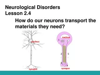
dendrite axon terminal postsynaptic neuron axon synapse How Do Neurons Communicate?
Neurotransmitter Classes • Acetylcholine • Amines (DA, NE, E, 5HT, histamine) • Amino acids (glutamate, GABA, glycine) • Purines (adenosine) • Gases (nitric oxide) • Neuropeptides (Sub P, endorphins, AII, oxytocin, many others)
Head Trauma / Bleeds • Focal: localized • Polar: acceleration-deceleration • Diffuse: widespread disruption
Determinants of Intracranial Pressure • Three space occupying components • Brain • CSF • Blood • Compensation for Increased ICP • CSF shunt to spinal cord • Hyperventilation leading to vasoconstriction
Causes of Increased ICP • Brain infection • Rupture of blood vessels • Hydrocephalus • F & E imbalances • Head Injury – most common
Types of Injury • Primary injury • Secondary injury
Ischemia ATP deficiency Release of glutamate Na+, Ca++ in cell “excitotoxin” Activation of phospholipases mitochondria dysfunction cell damage vasospasm platelet plug free radicals prostaglandins thromboxanes Pathophysiology of Secondary Injury
Compensation for Increased ICP Brain Swelling ICP CSF shunted to spinal cord Hyperventilation PaCO2 CSF in brain ventricles Cerebral vasoconstriction ICP Blood in brain ICP
Mild to moderate Moderate to severe Severe Headache, LOC, projectile vomiting, localized pain, decorticate posturingPupil changes, hyperventilation, decerebrate posturing, seizuresLoss of respiratory control, apnea Progression of S/S of Increasing ICP
Severe Severe Respiratory arrestFlaccidityIschemic responseBrain deathNo spontaneous respirations/3 minutesFixed pupilsFlat EEG Progression of S/S of Increasing ICP
Ischemic Response “Cushing’s Reflex” • Increased blood pressure • Wide pulse pressure • Decreased heart rate • Loss of respirations
Assessment of Brain Function • Level of Consciousness: ABCs • Manifestations of increased ICP • headache, vomiting, pupil reactivity • Glasgow Coma Scale • Eye Opening • Best Motor Response • Verbal Response • CT scan
General Therapy for Increased ICP • Elevate HOB • Diuretics • Sedation • Hyperventilation • Decompression
Classification of Head Injury • Concussion • Contusion • Brainstem Contusion • Hemorrhage * Epidural * Subdural - acute - subacute/chronic
epidural bleed subarachnoid bleed skull subdural bleed dura arachnoid Intracranial Bleeds
CVA: Stroke • Thrombotic • atherosclerosis, assess carotids > age 50 • Embolic • atrial fibrillation, valvular disease, hyper- coagulable states • Hemorrhagic • structural anomalies • hypertension
Stages of Thrombotic Stroke • Transient ischemic attacks (TIAs) • Stroke in evolution • Completed stroke
Manifestations of Stroke • Acute • focal neurological signs • may rapidly change (evolve) • depends greatly on area of brain damage • Transient Ischemic Attack (TIA) • signs and symptoms resolve quickly • no permanent loss of function
Stroke: Ischemic vs Hemorrhagic? • TIA: give ASA refer for carotid assessment • Stroke: Get CT scan immediately • Ischemic: evaluate for tPA (within 3 hours) • embolic and thrombotic • Hemorrhagic: Neurosurgical consult
Chronic Manifestations of Stroke • Contralateral hemiplegia • Ptosis • Homonymous hemianopsia • Neglect • Aphasia • Loss of bowel and bladder control • Emotional Instability
Homonymous Hemianopsia right visual field left visual field area of stroke damage left visual field blindness
General Therapy for CVA • Get to a Brain Trauma Center • Prevention • Manage high blood pressure • Anticoagulation • Rehabilitation
Alzheimer Disease • Dementia (deterioration of mentation) • about 70% Alzheimer type • others are multi-infarct type (vascular) • Manifestations (JAMICO) • judgment -confusion • affect -orientation • Memory • Intellect
Pathology of Alzheimer Disease • Genetics VS Environment • Apo-E gene • toxins, viruses, aluminum • Pathological Findings (at autopsy) • amyloid plaques • neurofibrillary tangles • cerebral atrophy and large ventricles
Alzheimer Disease • Diagnosis of Exclusion • rule out other, potentially treatable causes • MRI • brain atrophy, enlarged ventricles • Poor mental function • Mini Mental State Exam
Partial Simple (no LOC) Complex ( LOC) Secondarily generalized Generalized Absence (Petit Mal) Tonic-Clonic (Grand Mal) Seizures
Upper vs Lower Motorneuron UMN LMN Reflexes Increased Decreased Atrophy No Yes Muscle tone Spastic Flaccid Fasciculations No Yes
Upper Motor Neuron Disorders • Stroke/Head Injury • Cerebral Palsy • Huntington’s Chorea • Parkinson’s Disease
Localization of Motor Dysfunction • Reflexes • Deep tendon reflexes (cord reflexes) • Babinski (corticospinal tract) • Strength • focal vs general • ipsilateral vs contralateral • spasticity vs flaccidity
Parkinson Disease • Etiology • unknown, possibly neurotoxin • some suspect pesticide exposure • MPTP cases of Parkinson-like syndrome • Pathogenesis • Low dopamine level in basal ganglia • Excessive action of acetylcholine • Disease process is progressive
Manifestations of Parkinson Disease • Classic Triad (unilateral --> bilateral) • Akinesia • Rigidity • Resting tremor • Associated Manifestations • Propulsive gait - Poor speech quality • Masklike face - 30-50% have dementia • Drooling
Management of Parkinson Disease • Drug Therapy is controversial • Restore Dopamine / Ach balance • MAOI (selegiline) • Amantadine (Symmetrel) • Levodopa, carbidopa (Sinemet) • anticholinergics (Cogentin, Artane) • Surgical Techniques • adrenal medulla tissue transplants
Brainstem and Spinal Cord Disorders • Multiple Sclerosis • Poliomyelitis • Spinal Cord Injury
Multiple Sclerosis • Etiology • Autoimmune attack on CNS myelin • Pathogenesis • Immune injury to myelinated neurons • Sclerotic plaques noted on MRI • Demyelination disturbs neuron conduction • Extremely variable course and presentation
Presentation of MS • Usually relapsing remitting pattern • paresthesias • gait disturbance • leg weakness • vision loss (optic neuritis) • double vision • arm weakness • vertigo
Diagnosis and Treatment • Suspect with episodic neurologic deficits in 20-40 age group especially Northern European • MRI lesion is diagnostic • Treatment: symptoms • Beta interferon may decrease frequency of attacks • Immune suppression
Transection of Spinal Cord • Spinal Shock (lasts 2-8 weeks) • loss of spinal cord reflexes below injury • flaccidity • decreased vascular tone - hypotension • atony of bowel and bladder • Autonomic Dysreflexia • reflex activation of sympathetic neurons below level of injury
Autonomic Dysreflexia stimulus (full bladder) Reflex vasoconstriction below level of injury Increased blood pressure Can’t get signal to vessels below injury x Baroreceptor Response hypertension vasodilate above SCI bradycardia
transection of lateral cord Contralateral motor? sensory? Ipsilateral motor? sensory? Q: What Pattern of Sensory-Motor Impairment Would Occur?
Lower Motor Neuron Disorders • Bell’s PalsyGuillian Barre’ Syndrome
Guillain Barre’ Syndrome • Most common cause of acute flaccid paralysis • Presentation: Back leg pain progressing to weakness • decreased DTRs • Hx viral infection esp. mono preceding • decreased nerve conduction velocity • Hospitalize, plasmapheresis, IgG
Disorder of Neuromuscular Junction • Myasthenia Gravis • 80%-90% have anti-receptor antibodies • 75% have abnormal thymus Y Y Y
Myasthenia Gravis • Presentation: NM fatigue which worsens with activity: eye droop, diplopia, head droop, jaw dropping • No loss of reflexes, no change in sensation • Respond to edrophonium (fast acting anticholinesterase)