Download
1 / 29
390 likes | 708 Views
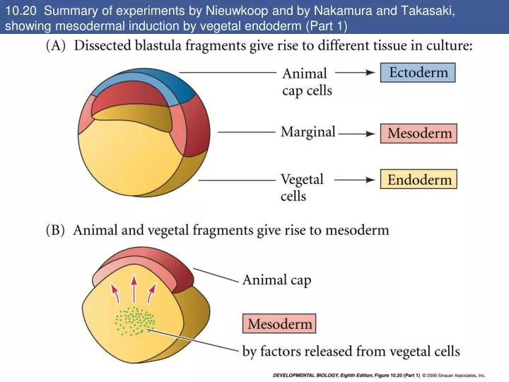
10.20 Summary of experiments by Nieuwkoop and by Nakamura and Takasaki, showing mesodermal induction by vegetal endoderm (Part 1). 10.20 Summary of experiments by Nieuwkoop and by Nakamura and Takasaki, showing mesodermal induction by vegetal endoderm (Part 2).

E N D
10.20 Summary of experiments by Nieuwkoop and by Nakamura and Takasaki, showing mesodermal induction by vegetal endoderm (Part 1)
10.20 Summary of experiments by Nieuwkoop and by Nakamura and Takasaki, showing mesodermal induction by vegetal endoderm (Part 2)
10.21 Experiments on 64-cell amphibian embryos demonstrated that the vegetal cells underlying the prospective dorsal blastopore lip region are responsible for causing the initiation of gastrulation
10.22 The regional specificity of mesoderm induction can be demonstrated by recombining blastomeres of 32-cell Xenopus embryos
10.23 The role of Wnt pathway proteins in dorsal-ventral axis specification • Early 2 cell stage; β-catenin in orange • Dorsal side of a presumptive blastula and nuclear β-catenin • No β-catenin in the ventral side • β-catenin dorsal localization through gastrula stage
10.24 Model of the mechanism by which the Disheveled protein stabilizes -catenin in the dorsal portion of the amphibian egg (Part 1)
10.24 Model of the mechanism by which the Disheveled protein stabilizes -catenin in the dorsal portion of the amphibian egg (Part 2)
10.25 Summary of events hypothesized to bring about the induction of the organizer in the dorsal mesoderm Goosecoid appears to be essential for specifying the dorsal mesoderm. Goosecoid expression occurs when there is a synergism between these proteins and TGF-β signal secreted by vegetal cells.
10.26 Model for mesoderm induction and organizer formation by the interaction of b-catenin and TGF-b proteins Nodal related gene Ventral and lateral mesoderm
Functions of the organizer (page 312) • While the Nieuwkoop center cells remain endodermal, the cells of the organizer become the dorsal mesoderm and migrate underneath the dorsal ectoderm. • Once the dorsal portion of the embryo is established, the movement of the involuting mesoderm establishes the anterior-posterior axis. • The endomesoderm that migrates first over the dorsal blastopore lip give rise to the anterior structures. • The mesoderm migrating over the lateral and ventral lips forms the posterior structures.
Induce the forebrain and midbrain • The organizer contribute to four cell types: • Pharyngeal endoderm • 2) Head mesoderm (prechordal plate) • 3) Dorsal mesoderm (primarily the notochord)- induces the hindbrain and trunk • 4) Dorsal blastopore lip- forms the dorsal mesoderm and eventually becomes the chordaneural hinge that induces the tip of the tail
10.27 Ability of goosecoid mRNA to induce a new axis The Nieuwkoop center activates the goosecoid gene in the organizer tissues. • Gastrula, one blastopore lip • Gastrula, two blastopore lips, • goosecoid was injected C) Goosecoid injected, 2 axes and controls D) Twinned embryo produced by goosecoid injection
10.28 Neural structures induced in presumptive ectoderm by newt dorsal lip tissue, separated from the ectoderm by a nucleopore filter with an average pore diameter of 0.05 mm • The epidermis that is induced to form, not the neural tissue. • The ectoderm is induced to become epidermal tissue by binding bone BMPs. • The nervous system forms from that region of the ectoderm that is “protected” from epidermal induction. • The “default fate” of the ectoderm is to become neural tissue; • Certain parts of the embryo induce the ectoderm to become epidermal tissue by secreting BMPs
10.29 Rescue of dorsal structures by Noggin protein • Injection of Noggin mRNA into 1-cell, UV-radiated embryos completely rescues dorsal development. • Noggin induces dorsal ectoderm to form neural tissue; • Noggin dorsalizes mesoderm cells that would otherwise contribute to the ventral mesoderm. • The development of dorsal structures is a dosage-dependent.
10.30 Localization of noggin mRNA in the organizer tissue, shown by in situ hybridization • Noggin mRNA is first localized in the dorsal blastopore lip region and then becomes expressed in the notochord. • Noggin binds to BMP4 and BMP2 and inhibits their binding to receptors. • At gastrulation at dorsal marginal zone • When cells involute in the dorsal blastopore lip • During convergent estension in the precursors of notochord • Extend beneath the ectoderm in the center of the embryo
10.31 Localization of chordin mRNA Chordin was found to be localized in the dorsal blastopore lip and later in the notochord. Of all organizer genes, chordin is the one most acutely activated by β- catenin. Chordin binds to BMP2 and BMP4 and prevents their complexing with their receptors.
Follistatin and BMPs • - Follistatin is also transcribed in the dorsal blastopore lip and notochord. • - Follistatin is an inhibitor of both activin and BMPs, causing ectoderm to become neural tissue. • In Xenopus, the epidermal inducers are BMPs (BMP4, BMP2, BMP7) and some relatives such as ADMP (anti-dorsalizing morphogenic protein). • BMP4 induced ectodermal cells to become epidermal.
10.32 Model for the action of the organizer Thus, the epidermis is instructed by BMP signaling, and the organizer works by blocking that BMP signal from reaching the ectoderm above it.
10.34 Regional specificity of induction can be demonstrated by implanting different regions (color) of the archenteron roof into early Triturus gastrulae
10.35 Regionally specific inducing action of the dorsal blastopore lip
10.36 Paracrine factor antagonists from the organizer are able to block specific paracrine factors to distinguish head from tail (Part 1)
10.36 Paracrine factor antagonists from the organizer are able to block specific paracrine factors to distinguish head from tail (Part 2)
10.37 Cerberus mRNA injected into a single D4 blastomere of a 32-cell Xenopus embryo induces head structures as well as a duplicated heart and liver Induction of the most anterior head structures could be accomplished by a secreted protein called Cerberus which promotes the formation of cement glands (the most anterior region of the tadpole ectoderm), eyes, and olfactory placodes. Cerberus mRNA injected in a vegetal ventral Xenopus blastomere originated head structures.
10.38 Xwnt8 is capable of ventralizing the mesoderm and preventing anterior head formation in the ectoderm • Frzb and Dickkopf are synthesized in the involuting endomesoderm (the most anterior portion of the organizer). • Frzd is a small, soluble form of Frizzled that can bind to Wnt. • - Dickkopf also appears to interact directly with the Wnt receptors, preventing Wnt signaling.
10.39 Insulin-like growth factors enhance anterior neural development IGFs are required for the formation of the anterior neural tube with its brain and sensory placodes. IGFs accumulate in the dorsal midline and are specially prominent in the anterior neural tube (A). B) Igf2 injected into ventral marginal zone blastomeres showing ectopic headlike structure. C) Control and IGF inhibitor
10.40 The Wnt signaling pathway and posteriorization of the neural tube (Part 1) The primary protein involved in posteriorizing the neural tube is thought to be a member of the Wnt family of paracrine factors, most likely Xwnt8. • It appears that a gradient of Wnt proteins is necessary for specifying the posterior region of the neural plate. • - In Xenopus, a gradient of Wnt signaling and β-catenin is higher in the posterior and absent in the anterior.
10.40 The Wnt signaling pathway and posteriorization of the neural tube (Part 2) • There appear to be two major gradients in the amphibian gastrula: • - BMP gradient that specifies the dorsal-ventral axis and • - Wnt gradient that specify the anterior-posterior axis
10.41 Model of organizer function and axis specification in the Xenopus gastrula
10.42 Pitx2 determines the direction of heart looping and gut coiling The embryo has a right-left axis. The crucial event in this axis formation is the expression of a nodal gene in the lateral plate mesoderm on the left side of the embryo. In Xenopus, this gene is Xnr1. It seems that the microtubules are involved in the establishment of the Xnr1 expression pattern. Vg1 seems to processed into its active form predominantly on the left hand side of the embryo. Vg1- Xnr1- activates pitx2 Pitx2 persists on the embryo’s left side as the head and gut develop, controlling their respective positions.