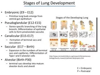
Download
1 / 34
430 likes | 965 Views
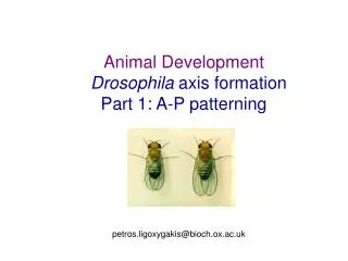
Drosophila embryonic Dorsal-Ventral patterning petros.ligoxygakis@bioch.ox.ac.uk. In this lecture. The origin of D-V asymmetry Maternal effect genes Dorsal/Ventral patterning

E N D
Drosophila embryonic Dorsal-Ventral patterning petros.ligoxygakis@bioch.ox.ac.uk
In this lecture • The origin of D-V asymmetry • Maternal effect genes • Dorsal/Ventral patterning • Multiple signalling pathways are involved in setting up D-V patterning. These pathways are used in different contexts during development and are highly conserved in different organisms.
The egg production line • Anterior Posterior • Young Old
As the ovarioles develop, follicle cells migrate over the oocyte and the nurse cells pump their contents into the oocyte, leaving only a remnant of the nurse cells behind at the anterior end. • A Drosophila egg is 400 mm long, 160 mm in diameter.
Early development in Drosophila After thirteen divisions membranes ingress from the cortex to enclose each nucleus, to form the cellular blastoderm.
Dorso-ventral axis formation • The egg is polarised on the DV axis (compare with animals where DV polarity is defined by the sperm entry point). • The ovary is not polarised on the DV axis, so how is the asymmetry established?
grk mRNA Grk protein stage 10 Grk protein gurken (a TGFb family protein) produced by the oocyte induces dorsal follicle cell fate, the first sign of D-V axis formation. Dorsal follicle cells stage 8 Ventral follicle cells • Step 1: Microtubule re-organisation. • Oocyte nucleus moves to an anterior position near the oocyte cell membrane. • grk mRNA is made in the nurse cells, transported to the anterior of the oocyte, then to the cortex overlying the oocyte nucleus, and anchored there. • Grk protein is synthesised, has limited diffusion, and signals to the follicle cells migrating overhead, which take on dorsal fates.
D/V patterning involves a Serine protease cascade • Share homology with trypsin-like family of extracellular serine proteases. • Typically secreted as inactive zymogen forms that are activated by proteolytic cleavage between N and C terminal domains. • Pre-activated forms of Snake and Easter lacking N-terminal sequences have been used to order Gastrulation defective, Snake and Easter in a cascade • In vitro Easter can cleave Spatzle to create active form in embryos • Snake and Easter zymogens are freely diffusible, therefore local activation is critical
Problem: Dorsal protein is the transcription factor that interprets the DV information in the egg. Dorsal as well as Toll (the receptor of the pathway) and its ligand Spatzle are found throughout the syncytial blastoderm, not just the ventral or dorsal side! How can Dorsal act as a morphogen and Toll signalling generate its gradient only in the ventral side? Solution: The critical step is the generation of the ventral signal by the only ventral-specific gene: Pipe. Translocation of Dorsal from the cytoplasm to the nuclei of the ventral cells occurs during the 14th cycle of cell division. Nuclei that take up Dorsal express ventralising genes and repress dorsalising genes. Question: What is the asymmetrical cue that leads to translocation of Dorsal into the nuclei of only ventral cells (Pipe target)?
Spn27A Easter Snake Gastrulation Defective Nudel Controlling the nuclear translocation of Dorsal CHORION PERIVITELLINE SPACE Spaetzle EMBRYO Windbeutel And pipe Dorsal Cactus Tube Toll Pelle MyD88 Plasma membrane Nuclear membrane FOLLICULAR CELLS
Organisational similarities of proteolytic cascades in development, coagulation and immunity
Generation of dorsal-ventral polarity In egg, after fertilisation. 7. Nudel and the Pipe target (factor x) interact to split the Gastrulation-deficient (Gd) protein. Nudel may determine the timing of this signal. 8. The activated Gd protein splits the Snake (Snk) protein, and activated Snk cleaves the Easter (Ea) protein. Gd, Snk and Ea are serine proteases 9. The activated Easter protein splits Spatzle; activated Spatzle binds to Toll receptor protein. 10. Toll activation activates Tube and Pelle, which phosphorylate the Cactus protein. Cactus is degraded, releasing it from Dorsal. 11. Dorsal protein enters the nucleus and ventralises the cell.
Searching for the target of Pipe Zhang et al (2009), Curr Biol 19, 1200-1205
Vitelline Membrane Like (VML) is a Pipe target and is localised at the anterior-lateral side of the oocyte
Genetic Interactions of vml with pipe Maternal Mutant Background Proportion of Embryos Exhibiting Denoted DV Phenotypes D0 D1 D2 D3 +/+; +/+; pipe7/pipe2 (n = 1681) 15.7% ± 0.9% 40.7% ± 1.2% 31.5% ± 1.1 12.1% ± 0.8% Vml/Vml; +/+; pipe7/pipe2 (n = 554) 89.5% ± 1.3% 10.3% ± 1.3% 0.2% ± 0.2% 0 Vml/+; +/+; pipe7/pipe2 (n = 327) 48.0% ± 2.8% 39.1% ± 2.7% 11.6% ± 1.8% 1.2% ± 0.6% gdVM90/+; +/+; pipe7/pipe2 (n = 208) 78.8% ± 2.8% 20.2% ± 2.8% 1.0% ± 0.7% 0.8%
Toll signalling Domain structure of Toll • Toll is a transmembrane protein found throughout the cell membrane of the egg that acts as a receptor for a localised external signal (Spatzle) • Toll membrane protein is activated by Spatzle on the ventral side of the embryo • In wild type embryos, amount of ligand is limited; in wild-type embryos Toll limits the diffusion of its own ligand by sequestering it as soon as it is produced. signal peptide (locates protein to membrane) intra-cellular domain (26% amino-acid identity with human interleukin-1 receptor transmembrane domain
Members of the Toll-like Receptor family TLRs are an evolutionary ancient and well conserved family of proteins Human TLR family consists of 10 members: TLR1-TLR10
Separation of dorsal and cactus proteins • Cactus binding to Dorsal protein inhibits Dorsal’s nuclear entry sequence, and thus Cactus sequesters Dorsal in the cytoplasm. Dorsal is a transcription factor Pelle protein kinase, probably through an intermediate, phosphorylates Cactus protein. P Cactus binds via ankyrin repeats Cactus degrades Dorsal protein can enter nucleus
D-V patterning so far… • D-V patterning is initially set up during oogenesis, via asymmetric mRNA localisation. • Extracellular proteolysis provides ligand ventrally for the ubiquitous receptor encoded by Toll. • Cactus proteolysis, releasing the morphogen Dorsal into ventral embryonic nuclei. • D-V patterning depends on cell-cell signalling rather than on localised determinants in the egg. • The signals controlling Dorsal access to nuclei use exclusively maternal products until the receptor coded by Toll is activated. • Dorsal and cactus then have both maternal and zygotic contributions • Transcriptional targets of Dorsal are zygotic.
Dorsal-ventral polarity • D-V axis is initially set up by maternal genes and depends on cell-cell signalling rather than on localised determinants in the egg. The role of Dorsal: • dorsal, which encodes a transcription factor that can both activate and repress gene expression, is the morphogenetic agent for D-V polarity. • Loss-of-function mutations in dorsal give rise to dorsalised embryos (the product of dorsal is needed for differentiation of ventral cells). • Different concentrations of Dorsal specify different patterns of gene transcription and consequently different fates for cells.
Differential nuclear localisation of dorsal protein regulates zygotic genes • Dorsal activates specific genes to give the mesodermal phenotype (nervecord, muscles etc.) and proper gastrulation. • twist, snail and rhomboid • Dorsal represses dorsalising genes • decapentaplegic (dpp) and zerknullt (zen)
How does Dorsal act as both a transcriptional activator and repressor? Dorsal twist • two types of complex - • activation • repression (low affinity binding site) DSP1 (HMG-box binding protein) TATA binding protein Dorsal (high affinity binding site) zen groucho (transcriptional repressor)
The gradient of the Dorsal protein and its interpretation (A) The concentration gradient of Dorsal protein in the nuclei of the blastoderm, as revealed by an antibody. (B) The interpretation of the Dorsal gradient by genes that demarcate the different dorsoventral territories; for simplicity, only two representative genes are shown. Subsequent processes will further subdivide these territories. The decapentaplegic (dpp) gene in particular codes for a secreted factor that will act as a local morphogen to control the detailed patterning of the ectoderm.
The gradient of the Dorsal protein and its interpretation (A) The concentration gradient of Dorsal protein in the nuclei of the blastoderm, as revealed by an antibody. (B) The interpretation of the Dorsal gradient by genes that demarcate the different dorsoventral territories; for simplicity, only two representative genes are shown. Subsequent processes will further subdivide these territories. The decapentaplegic (dpp) gene in particular codes for a secreted factor that will act as a local morphogen to control the detailed patterning of the ectoderm.
A dorsal-ventral gradient in Dpp is produced by the antagonistic activity of the short gastrulation protein (Sog). • The maternal gradient of dorsal protein in the nuclei represses dpp transcription ventrally but not dorsally. Sog is expresses in the ventral region of the embryo. Sog protein diffuses into the dorsal region and antagonizes the activity of Dpp protein, providing positional information in the dorsal region. Dpp Sog
Dorso-ventral patterning is controlled by the same genes in flies and frogs
Flies and frogs • Dorsal neural tube of the vertebrate and ventral nerve cord of the fly appear to be generated by the same set of instructions. fly frog Tolloid Xolloid Sog Chordin Dpp BMP-4
Can the fly sog gene rescue ventralised Xenopus embryos? • sog and chordin can substitute for each other! Ventralised embryos partially rescued by injection of sog mRNA Ventralised embryos completely rescued by injection of sog mRNA Embryos completely ventralised by UV irradiation
Summary • Similar signal transduction pathways in all multicellular organisms. • Homologous pathways form basic infrastructure, but targets may vary. • Molecular pathways are “tool-kits” comprising versatile ligands and receptors and molecular switches including proteolysis and reversible protein phosphorylation. • Signalling pathways can be recruited for different purposes. • Drosophila development shows significant similarities with vertebrate developmental systems.
Reading List • Wolpert et al, Principles of Development • Belvin MP and Anderson KV (1996) A conserved signalling pathway: the Drosophila Toll-dorsal pathway. Ann Rev Cell Dev Biol 12, 393-416 • Moussian B and Roth S (2005) Dorsoventral axis formation in the Drosophila embryo-shaping and transducing a morphogen gradient. Curr Biol 15, R887-899 • Further reading: Zhang et al, 2009 Curr Biol 19, 1200-1205