Circulatory system
Circulatory system. BLOOD- Fluid Connective Tissue. Amount of Blood present in the body of an adult human being –about 5-5.4 liters (a bucketful) Structural Organization of Blood: The Human Blood is classified into two major components- a) Cellular b) fluid. Cellular Components of Blood.
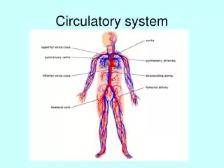

Circulatory system
E N D
Presentation Transcript
BLOOD- Fluid Connective Tissue • Amount of Blood present in the body of an adult human being –about 5-5.4 liters (a bucketful) • Structural Organization of Blood: • The Human Blood is classified into two major components- • a) Cellular b) fluid
Properties of RBCs • Formed in Adults- Red bone Marrow • Life cycle – 120 days • Destroyed-In liver • Mature RBCs in Mammals DO NOT have Mitochondria, Nucleus, Endoplasmic Reticulum. • Contains Pigment Hemoglobin
Leucocytes • Leucocytes- also called WBCs. • round, nucleated . • makes up the body defence system. Controls immunity by engulfing pathogens by phagocytosis.
Thrombocytes- also called Platelets These are cells which help in the process of formation of blood clot (Thrombus) at the site of injury
Mechanism of blood clotting • Ruptured platelets releasethromboplastinalso called thrombokinase enzyme. • Thromboplastin acts upon inactive prothrombin from ruptures RBCs and converts it into activated Thrombin, with Vitamin K and calcium ions acting as co-factors. • Thrombin acts upon soluble plasma protein fibrinogen and converts it into an insoluble mesh work of fibrin. • The wound is plugged • A straw coloured liquid oozes out of the clot. This is called Serum. Serum = Plasma – Fibrinogen.
BLOOD • Fluid Components: • also called plasma • contains proteins (fibrinogen is to be mentioned) • 92% is water. It also contains salts, dissolved nutrients, hormones, enzymes, etc
Structure of the Heart • Location of the heart – Thoracic cavity. (Chest cavity) • Protection of the heart – Bony – (Rib Cage) and membranous (pericardium) • Heart Muscles: 1. The human heart is a muscular pump. 2. Cardiac muscle is also involuntary. So functionally, cardiac muscle and smooth muscle are similar. 3. Cardiac muscle does have several unique features. Present in cardiac muscle are intercalated discs, which are connections between two adjacent cardiac cells. Intercalated discs help multiple cardiac muscle cells contract rapidly as a unit. This is important for the heart to function properly.
Structure of the Heart • Heart Chambers: 1. There are four chambers in the heart - two atria and two ventricles. The atria (one is called an atrium) are responsible for receiving blood from the veins leading to the heart. When they contract, they pump blood into the ventricles. 2. The muscle in the walls of the ventricles is much thicker than the atria as they need to push blood over a greater distance.
Structure of the Heart 3. Between the atria and the ventricles are valves, overlapping layers of tissue that allow blood to flow only in one direction.Valves are also present between the ventricles and the vessels leading from it. 4. The right side of the heart contains impure blood and the left side of the heart contains pure blood. The four chambers of the heart ensure that there is no mixing of pure and impure blood. This structure ensures efficient supply of oxygen to the body to meet with the high energy demands.
Cardiac cycle • The heart muscles are involuntary and under the control of a self excitatory nerve center situated at the roof of the right auricle called Sino-auricular node. This node is also called the pace maker or pace setter
Cardiac cycle • The cardiac cycle comprises of three stages: Auricular systole- a) Under the influence of an impulse from SAN the auricular chambers undergo systole. b) This causes the impure blood to enter from Rt. auricle to Rt. Ventricle via the tricuspid valve and pure blood from the lt. auricle to lt. ventricle through the Bicuspid or Mitral valve. c) These valves always open into the ventricles as their upturning into the auricles is prevented by chordae tendinae.
Cardiac cycle • Ventricular systole- • During this phase the Auriculo-ventricular node picks up the impulse from the SAN and sends it down the bundle of nerves called Bundle of His which divide into Purkinje fibres. • The Ventricles contract pushing the blood against the tricuspid and bicuspid valves which close to prevent back flow of blood. This generates the first sound of the heart beat- LUBB • Pure blood moves into the aorta, while impure blood enters into the Pulmonary artery through the semi-lunar valves.
Cardiac cycle • Joint Diastole • a) During this phase the auricles are in their relaxed state, while the ventricles begin to relax. • b) Due to differential pressure blood tends to flow back from The Aorta to the left Ventricle and from the Pulmonary Artery to the right Ventricle nut is prevented from flowing back as the semi-lunar valves close –generating the second sound of the heart beat –DUP
Double Circulation • During a Cardiac Cycle The blood flows circulates in two cycles.: • A) Between Heart and Lungs- Called Pulmonary Circulation. • B) Between Heart and the entire body – Called Systemic Circulation.
Hi! Just wanted to remind you that you must NOT forget to write the two terms- SYSTEMIC and PULMONARY While describing DOUBLE CIRCULATION.
Blood pressure-The ratio of systolic pressureto diastolic pressure during a Cardiac Cycle. Its numerical value is 120 / 80 mm of Hg. • The apparatus used to measure blood pressure is called Sphygmomanometer.
Hope you understood how I work !!! If you Still have doubts FEEL FREE to Get them cleared ANYTIME During school working hours.
Artery V/s Veins • Artery • Always carry blood away from the heart • Always carry oxygenated blood except pulmonary artery. • They have thick muscular walls • They have narrow lumen • They dilate and constrict with the heart beat- This is called pulse • They carry blood under pressure • They are deeply seated in the body. • Vein • Always carry blood towards the heart • Always carry de-oxygenated blood except pulmonary vein. • They have thin muscular walls • They have broad lumen • They do not dilate and constrict with the heart beat. • They do not carry blood under pressure. They have uni-directional valves at regular intervals. • They are superficially seated in the body.
LYMPH • Lymph is a fluid which is present in the lymphatic vessels. • This fluid is primarily the extra tissue fluid which is returned back into the venal blood. • It is similar to plasma except that it has a number of lymphocytes. • At places the lymph vessels get enlarged to form lymph nodes which are the store houses of lymphocytes. • Lymph contains tissue secretions like hormones, enzymes etc.
Functions of Lymph • The lymphatic system has three primary functions. First of all, it returns excess interstitial fluid to the blood. • The second function of the lymphatic system is the absorption of fats and fat-soluble vitamins from the digestive system and the subsequent transport of these substances to the venous circulation. • The third and probably most well known function of the lymphatic system is defense against invading microorganisms and disease.
Transport of Water and Minerals in Plants Ascent of sap- • Active uptake of ions from soil into roots • Endosmosis of water. • Generation of Root pressure • Transpiration Pull • Cohesive –Adhesive Forces
Ascent of sap- • Active uptake of Ions- The cells in the roots actively absorb ions from the soil solution to maintain the inner solution at a hypertonic level. This ensure that water always moves from the soil solution (hypotonic)into the solution inside the roots (Hypertonic) • Roots pressure – As the water moves from one cell to the next inside the root hairs and roots there develops a pressure, due to accumulation of salts at the basal cells of xylem called root pressure that pushes the water column into the xylem
Ascent of sap- • Root pressure alone cannot push the water column right up the tall trees. • Transpiration Pull. – as the wateris lost from the aerial parts of the trees as vapour called transpiration there develops a suction pull that pulls the entire water column upwards. This is called Transpiration Pull Theory – proposed by Jolly and Dixon
Ascent of sap- • Cohesion- Tension Theory- According to this theory there exists a force of attraction between water molecules (cohesive forces) and between water molecules and the walls of the xylem (adhesive forces). • These forces ensure the continuity of the water column and help in conduction of water – Ascent of sap.
Translocation of Food • Food is prepared in leaves in plants and are translocated by the phloem.The channels of transport are the sieve tubes. The force required for translocation is provided by the companion cells. • Food material move from the region of manufacture into the phloem by active process.
Translocation of Food • After entering the sieve tubes the nutrients being in high concentration exert an osmotic pressure which causes the entry of water into this region. • A high turgor pressure develops. • This pressure forces the nutrients to move towards the region with low turgor pressure.


