Download
1 / 25
250 likes | 391 Views
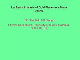
Ion Beam Analysis of Gold Flecks in a Foam Lattice F E Gauntlett, A S Clough Physics Department, University of Surrey, Guildford, GU2 7XH, UK. General Background

E N D
Ion Beam Analysis of Gold Flecks in a Foam Lattice F E Gauntlett, A S Clough Physics Department, University of Surrey, Guildford, GU2 7XH, UK
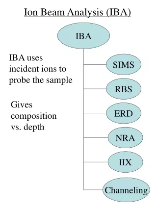
General Background • Clumpiness of the interstellar medium may play an important role in the transfer of infrared continuum radiation in star forming regions • Laboratory experiments have started, using high power laser facilities, to create clumpy media in the form of inhomogeneous plasmas to allow the testing of models of radiation transport Low density foam cylinder (1mm diameter 1mm) loaded with tiny gold spheres (~ 5 m diameter) Laser heated hohlraum
Ion Beam Characterisation of the gold-loaded foam • At Surrey we have been asked to measure: • the mass of the gold in the foam • the average size of the flecks • the uniformity of distribution of the flecks. • Facility: 2 MV Tandetron • Beamline: Scanning in vacuo proton microbeam • Techniques: Scanning Micro-PIXE • Scanning Micro-RBS • Sample: Gold loaded foam cylinder • Comparison sample: 2 m thick gold foil
Detectors At Ep ~ 4 MeV Au L X-rays (~ 10 keV) are produced in abundance but they are attenuated by ~ 35% in a 5 m diameter gold fleck. Au K X-rays have energies of 66 – 78 keV but their yield is low. However they are only attenuated by ~ 1%. Thus we need to detect both K X-rays to determine the overall quantity of gold present and L X-rays to determine from the attenuation the average size of the flecks. Hence we use eV Products CdTe detectors which are ~100% efficient between 10-70 keV. We also detect Backscattered protons with 100 sq mm ORTEC ULTRA detectors with a 300 m depletion layer. We use two detectors in both cases to obviate any instrumental asymmetries.
View of vertical sample holder , sample in carbon blocks and beam setting up plate
View of sample in the chamber through microscope at 135 to beam direction
RBS detector spectrum from foam containing gold flecks Carbon edge Oxygen edge Gold continuum
How do we explain the shape of the gold continuum? Assuming gold flecks are 5m in diameter and have a total mass of 15% of the foam mass – an upper estimate, the number of gold flecks in a cylinder is ~700 and the areal ratio (sum of cross sectional areas of gold spheres/area of foam cylinder) ~7x10⁻² i.e. 93% of the time protons in the beam incident on the foam go through the foam completely missing any gold flecks. It is likely that at most only 1 gold fleck is in the path of a proton. to detector 165O protons
Limiting Depth, DL At the highest backscatter detected energy (~4.05 MeV)only the front surface nuclei of the gold fleck at the left-hand edge will contribute. The Limiting Depth is when the backscatter energy from the front of a Au fleck there is equal to the backscatter energy from Oxygen at the front of the foam (~3.23 MeV). Contributions from different flecks at this backscatter energy are shown. At backscatter energies between 3.23 and 4.05 MeV fewer and fewer flecks contribute –giving the Au backscatter spectrum we observe
For 4 MeV protons DL~0.7mm. Thus to detect all the gold flecks in the foam to a depth of 1 mm with 4 MeV protons we detect X-rays. X-ray detector spectrum (Au L region) from foam with gold flecks
X-ray detector spectrum (Au K region) from foam with gold flecks
Au RBS top AuL PIXE right O RBS top AuK PIXE right Au RBS btm AuL PIXE left O RBS btm AuK PIXE left
Can we get an estimate of the gold mass? Backscatter spectra from the foam surrounds Top detector Bottom detector Counts per unit area
Taking a section of the carbon we measure carbon counts per unit area in a section of the spectrum NCS/ACS
Taking a separate run on the gold foil and adjacent carbon and software excluding the gold: Position of gold foil Position of Carbon Related Carbon BS spectrum From this we get NCF/ ACF
We then get an excellent measure of the beam charge ratio between the foam sample run and the foil run: QS/QF= (NCS/ ACS) /(NCF/ACF). We can use this to relate the Au K X-ray scatters from the Au flecks to those from the foil: NKS/NKF = (QS/QF)(MAu/MAuF)(d/d)K(ES)/(d/d)K(EF) whereMAuF is the mass of gold foil included in the scan and the differential cross-sections are evaluated at the mean proton energies ES, EFin the sample and foil respectively. We find MAu= 3.2 0.4 g
For the L X-rays: NLS/NLF = ((QS/QF)(MAu/MAuF)(d/d)L(ES)/(d/d)L(EF) exp((-/)SAul) / exp((-/)SAu (tF/2)) From this, using the gold mass MAu= 3.2 0.4 g, we can find a characteristic attenuation length l for X-rays in gold. This can be related to the gold diameter D, using an expression derived by Dirac, for the mean chord length in any one direction in a sphere: D = 3 l We find D = 5.5 0.6 m
An alternative technique is to measure the mass of gold flecks in foam is X-ray-fluorescence using a rhodium target x-ray tube. However this can only produce L X-rays from Au, eliminating the technique as a candidate for measuring 5 m diameter flecks. • So, to check our technique, we did measurements on a foam sample containing a similar mass of gold but having fleck diameters of ~0.5 ms. • In these flecks L X-ray attenuation is of order 1%. • From XRF measurements the gold mass is: MAu = 4.1 0.8 g • From our K X-ray measurements we find: MAu = 3.8 0.7 g • From our L X-ray measurements we find: MAu = 4.4 0.5 g All three measurements are compatible.
Another interesting feature of the measurement on the 0.5 m flecks is the BS spectrum – which, at the high backscatter end, looks like one you would expect from a low density gold film.
Estimate of mass from RBS Measurements Because of energy loss in the polyimide sleeve the gold backscatter spectrum above the Oxygen edge is representative of only a fraction of the cylinder area. From this though we can calculate the gold density. Assuming a uniform distribution of the gold we can then infer the mass by multiplying by the volume. It is 50.5 g.