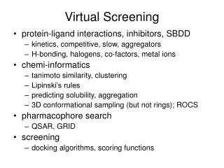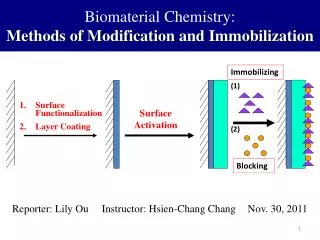
Virtual Screening Methods for Biomaterial Surface Functionalization
Spinal Progenitor Differentiation within Hyaluronic Acid Based Hydrogels for Spinal Cord Regeneration Sydney A. Geissler , Christine E. Schmidt Dept. of Biomedical Engineering, University of Texas, Austin. Sydney Geissler BME 4.202j sydneygeissler@mail.utexas.edu. Abstract.

Virtual Screening Methods for Biomaterial Surface Functionalization
E N D
Presentation Transcript
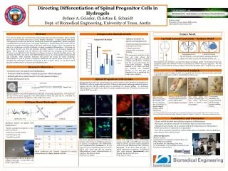
Spinal Progenitor Differentiation within Hyaluronic Acid Based Hydrogels for Spinal Cord Regeneration Sydney A. Geissler, Christine E. Schmidt Dept. of Biomedical Engineering, University of Texas, Austin Sydney Geissler BME 4.202j sydneygeissler@mail.utexas.edu Abstract Behavioral Analysis Spinal Progenitor Cells in Gels To determine extent of injury as well as to quantify the recovery, behavioral tests, previously optimized in collaboration with Dr. Tim Schallert, will be used. Over 200,000 people are currently living with paralysis due to spinal cord injury. Many of these injuries are sport related and occur in young, healthy individuals. A large problem with spinal cord regeneration is the scar that forms, blocking the axons from regenerating. Spinal progenitor implantation has been shown to encourage regeneration. When lacking a 3D construct, these cells tend to migrate toward the edges of the injury and become inactive. Here, I examined the effects of an HA hydrogel on spinal progenitor regeneration. Previously we have shown that hyaluronic acid (HA) can limit the astrocyte activation critical to scar formation around the injury. HA in generally non cell adhesive and would thus not encourage axonal regrowth, therefore I modified HA with laminin and collagen to form an injectable gelling system. By providing cells with a 3D matrix, their differentiation in an in vivo environment can be predicted and controlled. These injectable hydrogels will provide the matrix for spinal progenitor differentiation as well as provide a supportive scaffold for the injured spinal cord in vivo. By matching the mechanical properties of the gels to that of native tissue, we can direct spinal progenitor differentiation to neurons and oligodendrocytes. Research Objectives • Optimize matrix for spinal cord implantation. • Match mechanical properties to fetal spinal cord tissue. • Determine crosslink density • Determine pore size • Degradation profile • Optimize matrix for spinal cord implantation • Determine differentiability of spinal progenitors within hydrogels • Implant gels into a rodent model of cervical spinal cord injury • Assess functional recovery • Verify behavioral tests are translatable to contusion injury • Assess functional recovery of injured animals over 12 weeks • Tract tracing of corticospinal tract • Histological analysis of spinal cord injury Column 1 shows GFAP (glial) stain, column 2 shows β-III-tubulin (neuron) stain and column 3 is the overlay with DAPI (nuclear) stain. a. Spinal progenitors in collagen gels differentiate into astrocytes(scar forming glial cells, arrows). b. With HA and laminin added, spinal progenitors exhibit some differentiation into neurons (arrowheads). Scale bars are 50µm. Spinal Cord Spinal progenitor cells were isolated from E 12.5 mouse embryos and cultured as neurospheres in DMEM/F12 with N2 supplement and bFGF. Cells were dissociated and encapsulated into gels by mixing cells into the gel solution prior to incubation for thermal gelling. To encourage differentiation, cells in gels were cultured in media supplemented with 1% fetal bovine serum and without bFGF for 14 days. In collagen gels, cells differentiated into astrocytes. In HA-laminin-collagen gels cells differentiated into a mixed population of neurons and astrocytes. Discussion Virtual Screening Methods for Biomaterial Surface Functionalization • Currently there is no treatment for spinal cord injury • Research is needed to diminish scar formation and encourage regeneration • A 3D scaffold can provide structural support to the injured area • Spinal progenitor cells can encourage functional recovery • The 3D scaffold described here will direct progenitor cell differentiation • The addition of adhesive molecules may allow axonal regeneration • Behavioral tests to describe functional recovery are needed Hyaluronic Acid Based Hydrogels Spinal progenitor cells can be grown in collagen gels (left) and HA-laminin-collagen gels (right). The elastic compressive modulus of the two types of gels can be adjusted by changing the collagen content to mimic the native tissue. Lateral Hemisection Intact Spinal Cord Cervical Lateral Hemisection – Rodent Model + + 60% of spinal cord injuries are cervical injuries. By developing a rodent model for cervical spinal cord injury, we can better model the injuries and regeneration that may occur after injury. Hyaluronic Acid Laminin Collagen Acknowledgements: Mission Connect Lab Members: Dr. ZinKhaing (Post Doc), Michelle Jiang Dr. Tim Schallert (UT Psychology department and Institute for Neuroscience) The model we use currently is a transection model. We recently received an equipment grant from Mission Connect to purchase an Infinite Horizons Impacter. We will develop a cervical lateral contusion injury. Using the injectable gels I am developing, the cells and gels will be injected into the cavity that forms after injury. Astrocytes Oligodendrocytes Neurons Previously, we developed a transection model of cervical lateral hemisection spinal cord injury at C3/C4 in Sprague-Dawley rats. Spinal progenitor cells will be implanted into the gels to encourage regeneration. Previously our lab has shown neural progenitor cell differentiation within HA gels can be controlled by matching the mechanical properties to that of the native tissue. After cervical spinal cord injury, the cylinder test shows unilateral use of forelimbs for weight support during vertical exploration. The postural instability test reveals unilateral deficits during forward walking motion. The pasta eating test allows quantification of fine motor movement deficit as related to time to eat pasta. Vibrissae elicited placing reveals unilateral sensory motor deficits after cervical spinal cord injury