Download
1 / 36
510 likes | 2.53k Views
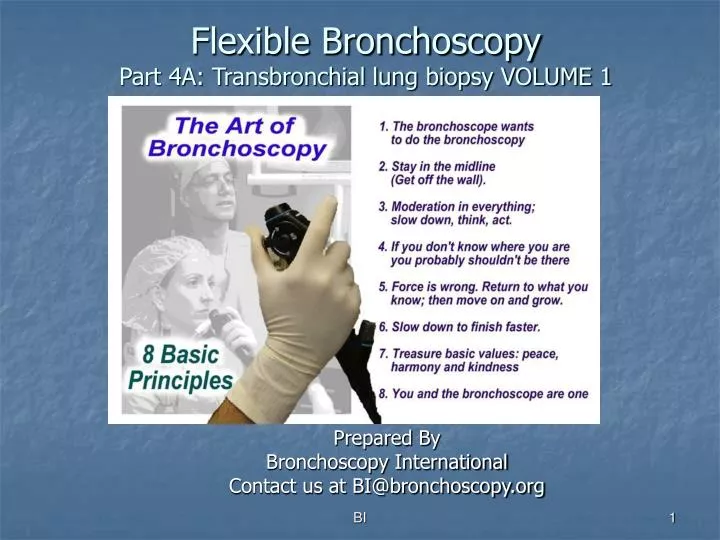
Prepared By Bronchoscopy International Contact us at BI@bronchoscopy.org. Flexible Bronchoscopy Part 4A: Transbronchial lung biopsy VOLUME 1. Strategy and Planning Execution. Transbronchial lung biopsy (TBLB). Prepared and distributed by Bronchoscopy International. History.

E N D
Prepared By Bronchoscopy International Contact us at BI@bronchoscopy.org Flexible BronchoscopyPart 4A: Transbronchial lung biopsy VOLUME 1 BI
Strategy and Planning Execution Transbronchial lung biopsy (TBLB) Prepared and distributed by Bronchoscopy International
History TBLB began to replace open lung biopsy in 1970’s in selected patients. • TBLB was originally considered very high risk • TBLB was originally performed in the operating theater . • TBLB performed by pulmonologists faced substantial opposition by surgeons. • TBLB was performed after endotracheal intubation. • Early history of TBLB was marked by frequent of bleeding, pneumothorax or respiratory failure. BI
TBLB today • Easily performed as outpatient procedure in a bronchoscopy suite. • Ideally performed using conscious sedation and topical anesthetic. • Fluoroscopy eliminates need for post-procedure chest radiograph and may increase patient safety. • Because most TBLB-related pneumothoraces occur during or immediately after TBLB, patients should probably be kept under observation for at least 1-2 hours after TBLB before being discharged home. Chest radiograph post-procedure should be obtained if symptoms are present. BI
Training is essential in order to • Learn proper techniques and indications • Avoid excessive procedure-related complications • Learn to treat procedure-related bleeding, pneumothorax, and respiratory failure • Learn to protect the equipment and avoid breaking the bronchoscope • avoid forced passage of the forceps through the scope at ANY time, especially if the scope is flexed • Avoid opening the forceps while it is inside the working channel of the bronchoscope. BI
When to perform TBLB • Usually, only after results from other bronchoscopic procedures such as BAL are negative, nondiagnostic, or considered not helpful depending on differential diagnosis. • Usually, only when results from TBLB will impact on disease management. • Usually, only when risks of the procedure have been satisfactorily understood by patient or family. BI
Contraindications to TBLB • Inadequate equipment • Insufficient training to assure efficacy and patient comfort and safety • Coagulopathy, patient on anticoagulation • Thrombocytopenia • Uremia (increases risks of bleeding) • Pulmonary hypertension (may increase bleeding risk) • Undue risk for respiratory failure or death in case of TBLB-related pneumothorax or bleeding • Examples: History of pneumonectomy, impending respiratory failure, poor lung function. BI
Presumed dangers of TBLB Biopsies of emphysematous lungs Biopsies around bullae and blebs Biopsies of stiff lungs of ILD Biopsies in vasculitis Biopsies of the middle lobe or lingula are adjacent to fissures Biopsies of superior segment of lower lobes are adjacent to fissures Avoid Non gravity dependent areas (anterior segment upper lobes) because bleeding may be difficult to control in these areas. Gough section: Upper lobe Emphysema BI
Complications of TBLB • Pneumothorax • Risk 1-4 % • Bleeding • 1.2 – 40% varies with studies and patient population. • Bleeding > 50 ml approximately 1-2% • Increased in uremia and immunocompromised patients • Death • Risk estimated at 0.04 -0.12 % BI
How does thatcompare to flexible bronchoscopy without TBLB ? • Bleeding in only 0.5 - 26 %. • Other adverse events include vaso-vagal reactions, reactions to anesthetics, bronchospasm, cardiac arrhythmias, and pneumothorax. • Mortality 0.01 - 0.05 %. BI
Risk of bleeding after Transbronchial lung biopsy • Perhaps a 45 % incidence in uremia (older studies). • < 15% incidence if PLT < 50,000. • Other concerns • Preprocedure laboratory studies often preferred • Importance of individualizing decisions based on H&P, Past medical History, Family History, and risk-benefit analysis. • One may consider stopping aspirin, other antiplatelet agents, and nonsteroidal anti-inflammatory drugs. One should definitely stop Plavix and anticoagulants (except subcutaneous Heparin used for prophylaxis). BI
Indications for TBLB • Diffuse and localized lung infiltrates suggestive of • Infectious lung disease (with negative or non helpful BAL) • Interstitial lung disease • Carcinoma or lymphoma • Pulmonary nodules and masses BI
Yield of TBLB • Nodules > 2 cm • 60% for lung cancer, 50% for metastatic disease • Inferior diagnosis in benign disease • AIDS • PCP • Mycobacteria • Kaposi • Kidney transplant and other immunocompromised hosts (poor for aspergillus, CMV, Mucor, Nocardia), but does add up to 10% yield to BAL ?) • Sarcoidosis: Usually > 80% • Interstitial lung disease: A diagnosis of fibrosis is Nonspecific and should be called NONDIAGNOSTIC BI
Yield in tumors • Primary tumor: yield > 60% • Metastases yield > 50% • Brushing increases yield • Lesions > 2.0 cm yield > 60 % • Lesions < 2.0 cm yield < 25% • Yields are lower in benign nodules BI
Yield in infiltrates yield is usually > 75 % for • Sarcoidosis • Alveolar proteinosis, • Lymphangitic carcinomatosis • Pneumoconiosis • PCP, CMV • Lung rejection • Bronchoalveolar cell carcinoma • Diffuse pulmonary lymphoma • Hypersensitivity pneumonitis BI
Diagnostic yield depends on • Bronchoscopist’s experience • Pathologist's experience • Predetermined criteria • if broad: yield > 72% • if narrow < 38% BI
predetermined criteria • Determine when results are accepted and acceptable. • Pathology interpretations may be difficult because of small specimens TBLB Forceps VATS BI
Number of specimens needed • PCP :at least 2 specimens if chest x-ray is Abnormal, and at least 4 specimens if chest x-ray is Normal (97% yield). • Sarcoid:Stage III, sensitivity increases with number (73-80% yield with at least 4 specimens, and increases further if endobronchial biopsies are done also. For Stage I Sarcoid, up to 10 specimens might be needed. • Transplant and lung rejection: Multiple specimens from multiple lobes are warranted. Yield > 60% for infection of rejection, but only 15 % for BO. Multiple specimens (> 6) are necessary. BI
Type of specimen : the Float sign • Float sign definition: Aerated lung floats, but nonaerated lung does not. • BUT, the float sign is not a reliable sign of representative alveolar and bronchiolar tissue. • Remember that increased number of biopsies increases diagnostic yield, but probably increases risk for complications with each biopsy. Partially aerated lung in patient with severe emphysema and iatrogenic pneumothorax BI
Size of specimens • Toothed (Alligator) forceps tear the lung more than cup forceps, and may cause more bleeding. • Large forceps obtain more tissue (more alveoli) than small forceps; frequency of bleeding is unchanged compared to smaller forceps. Am Rev Respir Dis 1993;148:1411-1413 Chest 1992;102:748-752. BI
Types of Forceps Cup Toothed BI
Fluoroscopy is often used for TBLB • Frequency of pneumothorax possibly increased if fluoroscopy is not used. • Avoids causing pleuritic chest pain with forceps. • Avoids need for post bronchoscopy radiograph because fluoroscopic examination at end of procedure determines presence or absence of TBLB-related pneumothorax. • Improves physician ease, comfort, and security • Used routinely by 75% of doctors in the USA. BI
Other advantages of fluoroscopy • Prevention of pneumothorax • Position of forceps in relation to pleura is visualized • Ability to obtain biopsies from localized infiltrate • Possibility to accelerate procedure • Avoid looking through the bronchoscope • Guidance possible using fluoroscopy image only, therefore scope can be wedged and forceps can be viewed using fluoroscopy only. BI
Fluoroscopy-assisted TBLB Position C-Arm first Test before starting bronchoscopy Be sure abnormalities can be seen on fluoroscopy Bronchoscopist should operate machine to avoid excess radiation Be certain that there is enough room in procedure area to assure patient safety in case of complications. Remove machine after biopsies Avoids need for post procedure radiograph BI
With fluoroscopy Forceps are easily inserted by the assistant into the bronchoscope if the scope is held “over the shoulder” Patients can be done supine or partially sitting BI
Fluoroscopy-assisted TBLB Once the scope is wedged, the Bronchoscopist watches the forceps using fluoroscopy only, and does not need to look through the bronchoscope until after all specimens are obtained In case of bleeding, the scope is kept wedged, suction is applied, and the patient is turned into the lateral safety position, bleeding side down. BI
Techniques of TBLB TBLB of the Right Lower lobe infiltrate, forceps open via lateral basal segment. TBLB of apical-posterior segment Left Upper Lobe, forceps still closed BI
Manipulating the Bronchoscope during TBLB • Wedge technique • Keeps scope in optimal position • Allows suction and tamponade in case of bleeding • Full view technique • Keeps segmental airways in view • Ability to better visualize bleeding if it occurs and to control patency of contra lateral lung • Ability to guide forceps into multiple specific segments BI
Full view and wedge techniques of TBLB Full view technique The scope is kept in a more proximal segmental bronchus Wedge technique The scope is wedged distally into the target subsegmental bronchus BI
Click to continue Wedge and nonwedge techniques of TBLB Click here to view video presentation nonwedge technique Click here to view video presentation Wedge technique BI
Touch and feel technique • Move forceps through working channel of scope. • As forceps becomes visible, begin fluoroscopy (intermittent rather than continuous decreases radiation exposure) • When forceps is at target position, open forceps and shake gently • Insert forceps a bit further until some resistance is felt • Ask the patient to raise a hand if pain is felt • This signals that the forceps is near the periphery of the lung and “touching” the pleura • Often used when fluoroscopy is NOT available • Increases the length of the procedure • Difficult if patients are well sedated • Close forceps, stop fluoroscopy, and withdraw forceps gently into working channel of bronchoscope. BI
Performing TBLB • When entering the apical segments of the upper lobes, keep the scope in the central airway and using only fluoroscopy to guide the forceps into the appropriate segment. • Seeing the target infiltrate in the retro cardiac and “sub diaphragmatic” regions. • Shaking the forceps if they don’t open immediately • If the scope is “over wedged”, pull forceps back slightly and bring the working channel into the midline and off the bronchial wall to make room for the forceps as it exits the working channel. • Change the angle of the forceps if they do not advance further into the periphery (forceps are probably caught on a spur) BI
Helpful hints for performing TBLB • Inform the patient that “there are no nerve endings in the airway, so the biopsy itself will not hurt”. • Use conscious sedation to improve patient comfort. • Forewarn the patient to “raise hand” if pain is felt at any time during the procedure. • Prefer biopsies from the lung periphery (as close to the pleura as possible) because bronchial vessels are smaller in the distal airways and forceps are most likely to “pinch” through bronchial mucosa to obtain representative tissue (contains alveoli and bronchioles) from lung parenchyma. • Avoid the lingula and right middle lobe because of proximity to fissures and risk of pneumothorax. BI
More Helpful hints for performing TBLB • When infiltrates are diffuse and involving the lower lobe, prefer biopsies from the lateral segment because fluoroscopically, the position of the forceps is true in relation to the chest wall. • Patient inhalation as the forceps is opened often allows the operator to advance the forceps further towards the periphery. • Keep the forceps open and advanced into the periphery for as short a time as possible, also keeping fluoroscopy time to a minimum (Usually < 30 seconds per biopsy). • Patient exhalation is followed by closure of the forceps. A quick and short tug is often followed by a patient inhalation. • By advancing the bronchoscope as the forceps is withdrawn, the scope is maintained in the wedge position. There is NO need to pull the forceps quickly up into the bronchoscope. BI
This presentation is part of a comprehensive curriculum for Flexible Bronchoscopy. Our goals are to help health care workers become better at what they do, and to decrease the burden of procedure-related training on patients. BI
Bronchoscopy International: Art of Bronchoscopy, an Electronic On-Line Multimedia Slide Presentation. http://www.Bronchoscopy.org/Art of Bronchoscopy/htm. Published 2007 (Please add “Date Accessed”). All efforts are made by Bronchoscopy International to maintain currency of online information. All published multimedia slide shows, streaming videos, and essays can be cited for reference as: Thank you BI