Download
1 / 77
800 likes | 1.15k Views
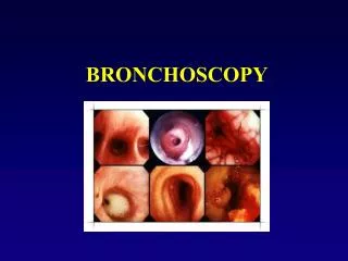
Basic Instrument s of Interventional Bronchoscopy. Ass. Prof. Sedat ALTIN Pulmonolog, interventional bronchoscopist. Rigid Bronchoscope.

E N D
Basic Instruments of Interventional Bronchoscopy Ass. Prof. Sedat ALTIN Pulmonolog, interventional bronchoscopist
Rigid Bronchoscope • The newer modifications in the rigid bronchoscope have established it as the ideal instrument for debulking of large tumors in the major airways, dilatation of tracheobronchial strictures, laser bronchoscopy, insertion of airway prostheses (stents), and extraction of tracheobronchial foreign bodies.
Fiberoptic Bronchoscope • Flexible bronchoscopes with a larger working channel enable the bronchoscopist to insert larger biopsy forceps, balloon catheters, laser fibers, and other instruments into the airways to obtain larger and better-quality biopsy specimens.
Videobronchoscope • A flexible bronchoscope equipped with a charge-coupled device at its distal tip. The bronchoscopic images are digitally captured and transmitted to a video processor for display on a television monitor. • The advantage is that the excellent images can be simultaneously visualized by many, making it an excellent tool for teaching purposes. The images can also be stored in several digital formats.
Videobronchoscope • The disadvantages include the added expense of purchasing video equipment and a computer terminal, and the larger working and storage space required. • The major drawback is the loss of ability to view the image through the headpiece of the flexible bronchoscope; the bronchoscopist has to depend on the video monitor to visualize bronchoscopic findings. The image on the monitor is only as good as the monitor.
EndoBronchialUltraSound (EBUS) • The major advantage of this technique is the ability to visualize, via ultrasound, theextra-airway structures that cannot be seen through the bronchoscope. The major technical problem is the inability to consistently provide the coupling of the ultrasound probe to thebronchial wall to generate meaningful images of the extrabronchial structures. To overcome this, flexible bronchoscopes are being fitted with water-inflatable balloons. This will permit constant 360-degree contact between the wall of the airway and the ultrasound probe. Preliminary studies have shown the ability to identify mediastinal structures including lymph nodes, great vessels, and esophagus .The identification of lymph nodes and their relation to airways may help improve diagnostic techniques such as BNA for the diagnosis and staging of thoracic tumors.
Fluorescence Bronchoscopy • When the normal bronchial mucosa is illuminated via the bronchoscope, a higher fluorescence is observed. Mucosa containing abnormal or malignant cells produces decreased autofluorescence. This phenomenon is used to detect mucosal changes suggestive of either premalignant or malignant lesions in the airway mucosa. Mucosal changes observed by routine (white-light) bronchoscopy can be compared with those observed via green-light bronchoscopy. Early reports show that this technique, when used as an adjunct to standard bronchoscopy, may enhance the ability to localize small neoplastic lesions, especially intraepithelial lesions.
Electromagnetic Guidance System A novel method for guiding transbronchial catheters or forceps is electromagnetic navigation. In comparison to fluoroscopy or CT scanning, electromagnetic navigation as a method not only has minimum technical and spatial requirements, it also indicates the position of the catheter in three dimensions without radiation exposure; all that it needs is the availability of a preprocedure CT data set.
Things to Consider • Personel • Scope of practice • Equipment • Space/unit • Your financial and practice environment
Minimal equipments • Rigid bronchoscopy system • Fiberoptic bronchoscopesandcoldlight source/video processor • Picture monitor • Forceps for biopsy, sitology brushes, transbronchial needlles, baskets for foreign bodies • Cleaning/disinfection equipments
Personel • A bronchoscopist alone is not enough • Nursing, ancillary help(minimal 2 nurses) • Anesthesiolog, technician • Good relationships with other services such as pathology, oncology and thoracic surgery • Requirements differ..
Equipment-Diagnostics • Basics • Good brochoscopes, at least 2 (videoscope) • Processors, screens etc…. • Image processing • Full range of forceps, brushes and TBNA needles
Equipment-Diagnostics • Advanced • AF if lung cancer detection program • EBUS • EM guidance system • Soon • NBI • Poss OCT
Equipment-Therapeutics • Basics • Therapeutic and thin flexible scopes • Choice of thermal ablation • Laser • ES/APC • Cryotherapy • Different diameter stents
Equipment-Therapeutics • Advanced • Rigid endoscopy with barrels, optics, camera and processor • PDT • Collection of silicone stents
interventional bronchoscopic procedures Complications, cautions, essential points
Procedures • Mechanical resection with rigid bronchoscopy • Dilatation • Laser (Nd-YAG, Nd-YAP), • Cryotherapy • Stent insertion • Photodinamic therapy • Brachitherapy • Argon Plasma Coagulation • Electrocautery
Conditions Required for Safety Endoscopic Treatment • Tumour must be accesible with the bronchoscope • Tumour must be spread restrictly in the bronchus and have to do not lymphangitic invasion • The lungs and the airways without stenosis must be functional • The performance of the patient must be enough good!!
Description of Common Practical Problems • Difficult airway management • Bleeding • Intubation • Indications and contraindications • Anesthesia and risk management
Which technical for which patient? • The type and the nature of stenosis • The localisation of the lesion • Available equipment must be preferred! • The experience of the physician • The condition of distal airways • The cost of the technics
Factors reducing the complications • Adequate equipment • Educated personnel • Sufficient sterilization • Good patient selection • Enough sedation, premedication and anesthesia • Follow-up after bronchoscopy and if need be therapy
Rigid Bronchoscopic Procedure-Related Adverse Events • poor insertion techniques, prolonged trauma of the larynx and vocal cords, or failure to heed the warnings of hypercapnia, hypoxemia, or hemodynamic instability • Airway wall perforation posterior wall of the trachea, subglottis, and median walls of the left and right main bronchi just below the carina Luxation or laceration of the vocal cords and arytenoids
Rigid Bronchoscopic Procedure-Related Adverse Events • Other complications can be avoided by a careful inspection of the mouth. Loose teeth should not be dislodged. The gums should not be traumatized, and the lips should not be injured. • Spinal cord injuries are possible in patients with cervical spine disease and severe osteoporosis. In selected instances, these diseases are contraindications to rigid bronchoscopic intubation.
Laser Bronchoscopy Attentions • FIO2 must be kept below 40% because of the risk of endobronchial fire • Avoid curare,pavulon because of post-operative respiratory depression. • Non flammable anaesthetic gases are mandatory. • An anesthesiologist experienced in the technique is important. • Oximetry monitoring is mandatory. • All persons in the room must wear protective glasses to avoid the risk of laser eye injury. • Plumbing- increased water for machine cooling • Electrical- special generator for high power needs. • RN Laser safety nurse • Laser operation-fiber bundle repair • Laser- $200,000maintainance- backup?
Laser Equipment • Dumon rigid laser bronchoscope with ventilating port, laser channel and suction channel. • Disposable large bore suction catheters. • Biopsy forceps with telescope. • Flexible bronchoscope. • Endobrochial balloon catheters in case of massive hemorrhage.
Laser Complications: • 1. Failure to achieve an adequate airway • 2. Hemorrhage usually mild and represents only a nuisance. • 3. Asphyxia • 4. Tracheoesophageal fistula can occur in LMB or tracheal lesions. • 5. Mediastinal emphysema, pneumothorax. • 6. Delayed hemorrhage (probably results from necrosis of tumor that had invaded a nearby pulmonary artery) • 7. Endobronchial fire • 8. Eye injury to the patient or OR staff
Cryo • In addition to the equipment needed for flexible or rigid bronchoscopy, dedicated operators need different probes depending on whether the cryotherapy is delivered through the rigid or flexible bronchoscope. Generally, the area of freezing is larger and the thawing quicker with the rigid probes. The gas most commonly used in cryotherapy and the gas most commercially available is nitrous oxide.
Cryotherapy • This technique is not indicated to achieve immediate debulking of an obstructive tumor. • The tumor will be first cored out mechanically with the tip of bronchoscope after coagulation, after first this step and inthe same session cryotherapy can be applied on the remaining infitrative part of the tumor. • Well vascularized tumor such as bronchial caecinomas, carcinoids,adenoid cystic carcinomas or granulomas • In situ or microinvasive carcinomas • CT is useful to remove many foreign bodies from the airways (pills, foods, clots, peanuts; not bones, metal,or teeth)
Cryotherapy • CT is not indicated in external compression of the bronchial tree, • CT is not indicated in benign strictures of the trachea or bronchi caused by fibroma, lipomas, or post-intubation stenosis • A transient fever immediately following cryotherapy. This fever can be prevented by corticosteroid administration given during the procedure • Airway sloughing material elimination after CT remains a problem. A bronchial toilet with a flexible fiberoptic bronchoscope is usually necessary 8-10 days after CT
Cryotherapy • The equipment is less expensive and easier to use than lasers. Subjective improvements have been observed in > 75% of patients with malignant airway lesions. • Complications are few and minor. One disadvantage is the longer duration of therapy required because of the need for frequent freeze-thaw cycles. Repeat bronchoscopy is needed for continued therapy in many patients.
Advantages of Cryotherapy • high penetration depth • no vaporization or carbonization • no smoke plume • fixation of liquids or tissue • can also be used to treat patients with cardiac pacemakers • no electrosurgical interference • no combustion risk • mobile unit
Advantages of Cryotherapy • Better control of depth effect • Can also be used in the area of coated stents • Does not harm cartilages • Less costs • approx. 7000 € /
APC & EC • a dedicated operator needs a high-frequency electrical generator in combination with insulated probes. Different types of probes in terms of shape as well as polarity (monopolar vs bipolar) are available. For patient and staff protection, proper insulation precautions need to be observed. Insulated flexible equipment is also available. For APC, a dedicated operator needs a special catheter allowing for the argon gas and the electrical current flow. This catheter is not used in electrocautery where there is direct tissue contact.
Argon Plasma Coagulation • The indication of APC is the same as that for laser: an obstructive endobronchial lesion of airway causing symptoms such as dyspnea, cough or pneumonia • The role of APC as a cure for early stage lung cancer is not yet fully established • In addition benign polyp removal and palliative care in malignant disease, it can also be used for debridement of granulation tissue around endobronchial stents. • APC has no role in removing a foreign body, mucous plug or clot. • Precautions: The power setting (<80W) and the application time (<5 sec)should minimize the risks and keeping the argon flow rate (<2 Lpm) should lessen the chance of gas embolism
Electrocauthery • Lesions considered suitable for the procedure were required to have < 50% luminal obstruction, a visualized size that was < 2 cm in its greatest dimension, limited vascularity, and an estimated procedure time of < 1 h.
Electrocautery • The diameter of the working channel of the scope is 2.6 mm, which allows the insertion of most therapeutic accessories. An electrosurgical unit was the power source for the procedure. This unit is approximately 1 cubic foot in volume and produces the three following current modes: cut, coagulate, and blend. The endobronchial accessories consisted of polypectomy snare, coagulation probe, forceps, and a cutting blade
Complications During Electrosurgery • Bleeding • Limited field of bronchoscope view • Transient desaturation • Excessive cough • Endobronchial fire • Electrical shock
Brachytherapy • Major complications include formation of fistulae between the airways and other thoracic structures in 6 to 8% of patients. Serious hemorrhage has been noted to occur more frequently in patients who receive high-dose radiation. The risk of massive hemoptysis increases dramatically when a fraction size of 15 Gy is used.
PDT • Complications from photodynamic therapy include sunburn involving skin exposed to bright light, hemoptysis, and expectoration of thick necrotic material.
Jean-François Dumon, MD, FCCP • ‘’Various types of airway stents available to treat airway stenoses. There is no ideal stent.’’
Stent indications Inoperable, symptomatic lung cancer• Primary airway tumours• Oesophageal cancer• Thyroid cancer• Head and Neck tumours• Metastases• Postintubation and idiopatic benign tracheal stenosis• Inflammatory lesions• Tracheobronchial malacia• Vascular compression
Stents • Complications seen with silicone stents include migration of stent and inspissation of thick mucous within the stent lumen. Metallic stents seem to promote growth of granulation tissue, which makes it difficult to remove and replace the stent. Uncovered metallic stents should not be inserted in patients with malignant airway lesions because the growth of cancer through the wire mesh negates the benefits of stent placement.
Bronchoscopic Needle Aspiration • Complications are rare and include pneumothorax and hemomediastinum. Serious bleeding is seldom encountered. More commonly, inadvertent passage of the needle through the wall of the working channel of the flexible bronchoscope leads to expensive damage to the inner lining of the bronchoscope.