Download
1 / 65
670 likes | 1.33k Views
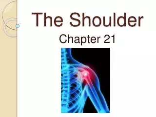
Chapter 22: The Shoulder Complex. The shoulder is an extremely complicated region of the body Joint which has a high degree of mobility but not without compromising stability

E N D
The shoulder is an extremely complicated region of the body • Joint which has a high degree of mobility but not without compromising stability • Involved in a variety of overhead activities relative to sport making it susceptible to a number of repetitive and overused type injuries
Functional Anatomy • Great degree of mobility w/ limited stability • Round humeral head that articulates w/ a flat glenoid • Ability of the rotator cuff & long head of the biceps provide dynamic stability • Supraspinatus compresses the head while the other rotator cuff muscles depress the humeral head during overhead motion • Integration of the capsule and rotator cuff • Muscle contractions dynamically control the capsule
Scapula stabilizing muscles and the relationship with the other joints of the shoulder complex and the glenohumeral joint • Scapulohumeral Rhythm • Movement of scapula relative to the humerus • Initial 30 degrees of glenohumeral abduction does not incorporate scapular motion (setting phase) • 30 to 90 degrees the scapula abducts and upwardly rotates 1 degree for every 2 degrees of humeral elevation • Above 90 degrees the scapula and humerus move in 1:1 ratio
Prevention of Shoulder Injuries • Proper physical conditioning is key • Develop body and specific regions relative to sport • Strengthen through a full ROM • Warm-up should be used before explosive arm movements are attempted • Contact and collision sport athletes should receive proper instruction on falling • Protective equipment • Mechanics versus overuse injuries
Assessment of the Shoulder Complex • History • What is the cause of pain? • Mechanism of injury? • Previous history? • Location, duration and intensity of pain? • Creptitus, numbness, distortion in temperature • Weakness or fatigue? • What provides relief?
Observation • Elevation or depression of shoulder tips • Position and shape of clavicle • Acromion process • Biceps and deltoid symmetry • Postural assessment (kyphosis, lordosis, shoulders) • Position of head and arms • Scapular elevation and symmetry • Scapular protraction or winging • Muscle symmetry • Scapulohumeral rhythm
Sternoclavicular joint Clavicular shaft Acromioclavicular joint Coracoid process Acromion process Humeral head Greater and lesser tuberosity Bicipital groove Spine of scapula Scapular vertebral border Scapular lateral border Scapular superior angle Scapular inferior angle Palpation - Bony
Sternoclavicular, acromioclavicular and coracoclavicular ligaments Rotator cuff muscles and tendons Subacromial bursa Sternocleidomastoid Biceps and tendon Coracoacromial ligament Glenohumeral joint capsule Deltoid Rhomboids Latissimus dorsi Serratus Anterior Levator scapulae Trapezius Supraspinatus Infraspinatus Teres major and minor Palpation - Soft Tissue
Special Tests • Active and Passive Range of Motion • Flexion, extension • Abduction and adduction • Internal and external rotation • Muscle Testing • Muscles of the shoulder and those that serve as scapula stabilizers • Test for Sternoclavicular Joint Instability • With athlete seated, pressure is applied to the SC joint anteriorly, superiorly and inferiorly to determine stability or pain associated w/ a joint sprain
Test for Acromioclavicular Joint Instability • Palpate for displacement of acromion and distal head of clavicle • Apply pressure in all 4 directions to determine stability • Tests for Glenohumeral Instability • Glenohumeral Translation - anterior and posterior stability
Anterior and Posterior Drawer tests • Sulcus test Clunk Test
Apprehension test and Relocation test • Apprehension test used for anterior glenohumeral instability (1) • Posterior instability apprehension test (2) • Relocation test uses external rotation and anterior pressure to allow for increased external rotation (3)
Test for Shoulder Impingement • Neer’s test and Hawkins-Kennedy test for impingement used to assess impingement of soft tissue structures • Positive test is indicated by pain and grimace
Tests for Supraspinatus Muscle Weakness • Drop Arm Test • Used to determine tears of rotator cuff (primarily the supraspinatus) • Athlete abducts shoulder and gradually lowers to starting position • Inability to lower arm slowly and controlled will indicate torn supraspinatus
Empty Can Test • 90 degrees of shoulder flexion, internal rotation and 30 degrees of horizontal abduction • Downward pressure is applied • Weakness and pain are assessed bilaterally
Test for Serratus Anterior Weakness • Wall push-up - looking for winging scapula • Could indicate injury to long thoracic nerve • Test for Biceps Irritation • Yergason’s test and Speed’s test utilized to determine pain and possible subluxation of biceps tendon • Ludington’s test used to assess possible rupture of biceps (feel for contraction while alternating contractions of each biceps)
Tests for Thoracic Outlet Compression Syndrome • Anterior scalene syndrome (Adson’s test) • Compression of subclavian artery by scalenes is assessed • Disappearance of pulse while athlete turns toward extended arm and takes a breath indicates a positive test • Costoclavicular syndrome test (Roo’s test) • Compression of artery between clavicle and first rib • Test is positive if after opening and closing hands for 3 minutes, strength or circulation decreases • Test is also positive if while in military brace position, head is turned in opposite direction and pulse disappears • Hyperabduction syndrome test (Allen test) • Used to assess if pressure from pectoralis minor is compressing brachial plexus and subclavian artery • Sensation Testing
Recognition and Management of Specific Injuries • Clavicular Fractures • Etiology • Fall on outstretched arm, fall on tip of shoulder or direct impact • Occur primarily in middle third (greenstick fracture often occurs in young athletes) • Signs and Symptoms • Generally presents w/ supporting of arm, head tilted towards injured side w/ chin turned away • Clavicle may appear lower • Palpation reveals pain, swelling, deformity and point tenderness
Clavicular Fractures (continued) • Management • Closed reduction - sling and swathe, immobilize w/ figure 8 brace for 6-8 weeks • Removal of brace should be followed w/ joint mobes, isometrics and use of a sling for 3-4 weeks
Scapular Fractures • Etiology • Result of direct impact or force transmitted up through humerus • Signs and Symptoms • Pain during shoulder movement as well as swelling and point tenderness • Management • Sling immediately and follow-up w/ X-ray • Use sling for 3 weeks w/ overhead strengthening beginning at week 1
Fractures of the Humerus • Etiology • Humeral shaft fractures occur as a result of a direct blow, or fall on outstretched arm • Proximal fractures occur due to direct blow, dislocation, fall on outstretched arm • May pose danger to nerve and blood supply • Epiphyseal fractures are more common in young athletes - occur due to direct blow or indirect blow travelling along long axis of humerus • Signs and Symptoms • Pain, swelling, point tenderness, decreased ROM • Management • Immediate application of splint, treat for shock and refer • Humeral fractures- remove from activity for 3-4 months • Proximal fracture - incapacitation 2-6 months • Epiphyseal fracture - quick healing - 3 weeks
Sternoclavicular Sprain • Etiology • Indirect force, blunt trauma (may cause displacement) • Signs and Symptoms • Grade 1 - pain and slight disability • Grade 2 - pain, subluxation w/ deformity, swelling and point tenderness and decreased ROM • Grade 3 - gross deformity (dislocation), pain, swelling, decreased ROM • Possibly life-threatening if dislocates posteriorly • Management • RICE, reduction if necessary • Immobilize for 3-5 weeks followed by graded reconditioning
Acromioclavicular Sprain • Etiology • Result of direct blow (from any direction), upward force from humerus, • Can be graded from 1-6 depending on severity • Signs and Symptoms • Grade 1 - point tenderness and pain w/ movement; no disruption of AC joint • Grade 2 - tear or rupture of AC ligament, partial displacement of lateral end of clavicle; pain, point tenderness and decreased ROM (abduction/adduction) • Grade 3 - Rupture of AC and CC ligaments • Grade 4 - posterior dislocation of clavicle
Signs and Symptoms • Grade 5 - loss of AC and CC ligaments; tearing of deltoid and trapezius attachments; gross deformity, severe pain, decreased ROM • Grade 6 - displacement of clavicle behind the coracobrachialis • Management • Ice, stabilization, referral to physician • Grades 1-3 (non-operative) will require 3-4 days and 2 weeks of immobilization respectively • Grades 4-6 will require surgery • Aggressive rehab is required w/ all grades • Joint mobilizations, flexibility exercises, & strengthening should occur immediately • Progress as athlete is able to tolerate w/out pain and swelling • Padding and protection may be required until pain-free ROM returns
Glenohumeral Joint Sprain • Etiology • Forced abduction and/or external rotation or a direct blow • Signs and Symptoms • Pain during movement especially when re-creating MOI • Decreased ROM and pain w/ palpation • Management • RICE for 24-48 hours; sling • After hemorrhaging subsides, cryotherapy, ultrasound and massage can be used along w/ passive and active exercise to regain full ROM • When full ROM achieved w/out pain, resistance exercises can be initiated • Must be aware of potential development of chronic conditions
Acute Subluxations and Dislocations • Etiology • Subluxation involves excessive translation of humeral head w/out complete separation from joint • Anterior dislocation is the result of an anterior force on the shoulder, forced abduction and external rotation • Posterior dislocation occurs due to forced adduction and internal rotation or falling on an extended and internally rotated shoulder • Signs and Symptoms • Anterior dislocation - flattened deltoid, prominent humeral head in axilla; arm carried in slight abduction and external rotation; moderate pain and disability
Signs and Symptoms • Posterior dislocation - severe pain and disability; arm carried in adduction and internal rotation; prominent acromion and coracoid process; limited external rotation and elevation • Management • RICE and reduction by a physician • Immobilize following reduction for 3 weeks • Perform isometrics while in sling • Progress to resistance exercises as pain allows • Return to play when athlete has regained 20% of body weight when tested for internal and external rotation • Protective bracing
Possible Complications of Shoulder Dislocations • Bankart lesion - permanent anterior defect of labrum • Hill Sachs lesion - caused by compression of cancellous bone against anterior glenoid rim creating a divot in the humeral head • SLAP lesion - defect in superior labrum that begins posteriorly and extends anteriorly impacting attachment of long head of biceps on labrum • Brachial nerves and vessels may be compromised • Rotator cuff injuries • Bicipital tendon subluxation and transverse ligament rupture
Chronic Recurrent Instabilities • Etiology • Traumatic, atraumatic, microtraumatic (repetitive use), congenital and neuromuscular • As supporting tissue become more lax, mobility increases resulting in damage to other soft tissue structures • Signs and Symptoms • Anterior - may have clicking or pain; complain of dead arm during cocking phase (when throwing); pain posteriorly; possible impingement; positive apprehension test • Posterior - possible impingement, loss of internal rotation; crepitation; increased laxity; pain anteriorly and posteriorly • Multidirectional - inferior laxity; positive sulcus sign; pain and clicking w/ arm at side; possible signs and symptoms associated w/ anterior and posterior instability
Chronic Recurrent Instabilities of the Shoulder • Management • Conservative treatment involves extensive strengthening (rotator cuff and scapula stabilizers) • Avoid joint mobilizations and flexibility exercises • Various harnesses and restraints can be used to limit motion • Surgical stabilization may be required to improve function and comfort • Strengthening should be continued for a reasonable time before surgery is opted for
Shoulder Impingement Syndrome • Etiology • Mechanical compression of supraspinatus tendon, subacromial bursa and long head of biceps tendon due to decreased space under coracoacromial arch • Seen in over head repetitive activities • Exacerbating factors - laxity and inflammation, postural mal-alignments • kyphotic posture, rounded shoulders • Signs and Symptoms • Diffuse pain, pain on palpation of subacromial space • Decreased strength of external rotators compared to internal rotators; tightness in posterior and inferior capsule • Positive impingement and empty can tests
Neer’s progressive stages of shoulder impingement • Stage I - result of supraspinatus or biceps tendon injury presenting w/ point tenderness, pain w/ abduction and resisted supination w/ external rotation; edema, thickening of rotator cuff and bursa • Occurs in athlete < 25 years old • Stage II - permanent thickening and fibrosis of supraspinatus and biceps tendon; presenting w/ aching during activity that worsens at night; May experience restricted arm motion • Stage III - history of shoulder problems and pain, tendon defect (3/8 “) or possible muscle tear and permanent scar tissue and thickening of rotator cuff • Athletes 25-40 years old • Stage IV- infraspinatus and supraspinatus wasting, pain during abduction, tendon defect greater than 3/8”, limited active and full passive ROM, weak resistive ROM and clavicle degeneration
Rotator cuff tear • Occurs near insertion on greater tuberosity • Partial or complete thickness tear • Full thickness tears usually occur in those athletes w/ a long history (generally does not occur in athlete under age 40) • Primary mechanism - acute trauma or impingement • Involve supraspinatus or rupture of other rotator cuff tendons • Management • Analgesics, electrical stimulation for pain, NSAID’s and ultrasound for inflammation • Restore appropriate mechanics and strengthen rotator cuff to depress and compress humeral head to restore space • Strengthen lower extremity and trunk to reduce stress on shoulder • Stage III and IV cases may require immobilization and rest and potentially surgery
Shoulder Bursitis • Etiology • Chronic inflammatory condition due to trauma or overuse - subacromial bursa • Fibrosis, fluid build-up resulting in constant inflammation • Signs and Symptoms • Pain w/ motion and tenderness during palpation in subacromial space; positive impingement tests • Management • Cold, ultrasound and NSAID’s to reduce inflammation • Remove mechanisms precipitating condition • Maintain full ROM to reduce chances of contractures and adhesions from forming
Frozen Shoulder (Adhesive Capsulitis) • Etiology • Contracted and thickened joint capsule w/ little synovial fluid • Chronic inflammation w/ contracted inelastic rotator cuff muscles • Generalized pain w/ motions (active and passive) resulting in resistance of movement • Signs and Symptoms • Pain in all directions both w/ active and passive motion • Management • Aggressive joint mobilizations and stretching of tight musculature • Electric stim for pain and ultrasound for deep heating
Thoracic Outlet Compression • Etiology • Compression of brachial plexus, subclavian artery and vein due to 1) decreased space between clavicle and first rib, 2) scalene compression, 3) compression by pec. minor, or 4) presence of cervical rib • Signs and Symptoms • Paresthesia and pain, sensation of cold, impaired circulation, muscle weakness, muscle atrophy and radial nerve palsy • Positive anterior scalene test, costoclavicular test and hyperabduction test • Management • Conservative treatment - correct anatomical condition through stretching (pec minor and scalenes) and strengthening (trapezius, rhomboids, serratus anterior, erector spinae)
Biceps Brachii Rupture • Etiology • Result of a powerful contraction • Generally occurs near origin of muscle at bicipital groove
Signs and Symptoms • Athlete hears a resounding snap and feels sudden and intense pain • Protruding bulge may appear near middle of biceps • Definite weakness with elbow flexion and supination • Management • Ice for hemorrhaging, place arm in sling and refer to athlete • Athletes will require surgery • Older individual will be able to rely on brachialis which serves as primary elbow flexor