Download
1 / 47
1.81k likes | 5.7k Views
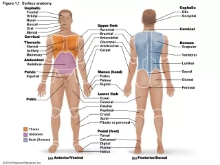
Surface Anatomy. Palpation – feeling internal structures through the skin “Living anatomy” – provides information about Palpation of arterial pulses Skeleton, muscles, and blood vessels Sounds of the heart and lungs Where to give injections. The Head.

E N D
Surface Anatomy • Palpation – feeling internal structures through the skin • “Living anatomy” – provides information about • Palpation of arterial pulses • Skeleton, muscles, and blood vessels • Sounds of the heart and lungs • Where to give injections
The Head • Cranium – selected structures felt through the skin • Superciliary arches • External occipital protuberance • Mastoid process • Temporalis muscle – at temple region • Frontalis muscle • Feel wrinkling of the forehead when eyebrows are raised
Surface Anatomy of Lateral Aspect of the Head Figure 11.26
The Head • Face – selected structures felt through the skin • Lacrimal fossa • Root and bridge of the nose • Auricle of the ear • Zygomatic arch • Masseter muscle • Mandible • Temporomandibular joint
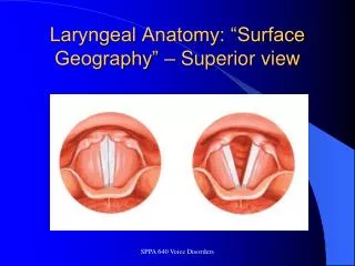
The Neck • Skeletal landmarks • Spinous processes of cervical vertebrae • C7 is particularly prominent (vertebra prominens) • Hyoid bone – in the anterior, superior neck • Laryngeal prominence – the “Adam’s Apple” • Cricoid cartilage – inferior to the laryngeal prominence • Jugular notch – depression in the superior part of the sternum
The Neck Figure 11.27
Muscles of the Neck • Sternocleidomastoid – most prominent neck muscle • Sternal head • Clavicular head • Deep to the sternocleidomastoid • Common carotid artery • Internal jugular vein • Trapezius – posterior aspect of the neck
Triangles of the Neck • The sternocleidomastoid muscles divide the neck • Anterior triangle • Posterior triangle Figure 11.28a
Triangles of the Neck Figure 11.28b
The Trunk • The trunk consists of the • Thorax • Abdomen • Pelvis and perineum
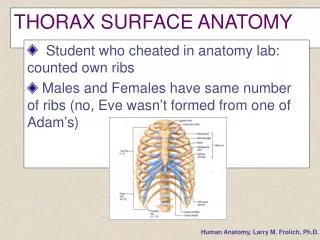
The Thorax • Sternum – portions felt through the skin • Manubrium, xiphoid process, and sternal angle • Midaxillary line – line from the center of the axilla onto the lateral thoracic wall • Midclavicular line – vertical line from midpoint of the clavicle to the groin
Muscles of the Thorax • Pectoralis major • Serratus anterior
The Anterior Thorax and Abdomen Figure 11.29
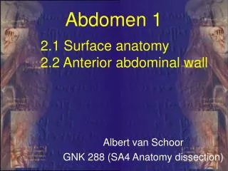
The Abdomen • Structures felt through the skin • Iliac crest • Anterior superior iliac spine • Inguinal ligament • Runs medially from anterior superior iliac spine to the pubic tubercle • Pubic crest
Inguinal Hernia Figure 11.30
Muscles and other Abdominal Surface Features • Linea alba • The “white line” extending from xiphoid process to the pubic symphysis • Rectus abdominis • Linea semilunaris – lateral margin of rectus abdominis
The Pelvis and Perineum • Four bony structures define the perineum • Pubic symphysis • Two ischial tuberosities • Coccyx
The Back • Posterior median furrow – vertical groove along the midline • Spinous processes of vertebrae • Spine of the scapula • Medial end is opposite T3 • Medial border of the scapula
The Back • Inferior angle of the scapula • Iliac crests • Supracristal line – intersects L4 • Sacrum – superior to cleft in the buttocks • Coccyx – posterior to the anus
Surface Anatomy of the Back Figure 11.31a
Muscles of the Back • Trapezius • Latissimus dorsi • Erector spinae
Surface Anatomy of the Back Figure 11.31b
Upper Limb and Shoulder • The Axilla • Base of the axilla – where armpit hair grows • Deep to the axilla – axillary lymph nodes and blood vessels • Anterior axillary fold – pectoralis major • Posterior axillary fold –latissimus dorsi and teres major
Shoulder and Arm Figure 11.32a, b
The Shoulder • Acromion – lateral end of the spine of the scapula • Acromioclavicular joint • Deltoid muscle • Covers the greater tubercle of the humerus Figure 11.33
The Arm • The region between the shoulder and elbow • Humerus • Palpated through skin along its entire length • Biceps brachii • Medial bicipital groove • The medial boundary of the biceps brachii • Triceps brachii
The Arm Figure 11.34
The Elbow • Lateral and medial epicondyles of the humerus • Ulnar nerve – “funny bone” runs across medial epicondyle • Olecranon process of the ulna • Cubital fossa – (antecubital fossa) • Forms anterior surface of forearm
The Anterior Surface of the Forearm and Fist Figure 11.35a
Forearm • Bones • Ulna – palpate entire length • Styloid process and head – distal end • Radius – partly covered in muscle • Head of the radius – proximal end • Styloid process – distal end
Muscles of the Forearm • Flexor muscles – anterior forearm • Flexor carpi radialis • Palmaris longus • This muscle is absent in about 30% of people • Extensor muscles – posterior forearm
Other Structures of the Forearm • Anatomical snuff box bordered by • Extensor pollicis brevis • Extensor pollicis longus
The Hand • Dorsum of hand • Dorsal venous network • Tendons of extensor digitorum • Palmar surface • Thenar eminence (not labeled on figure 11.35b) • Hypothenar eminence (see figure 11.17a–c ) • Pisiform bone
The Dorsum of the Hand Figure 11.36
Gluteal Region • Iliac crests • Posterior superior iliac spine • Sacroiliac joint • Prominences of the buttocks • “Cheeks” of the buttocks • Formed from subcutaneous fat and the gluteal muscles
The Gluteal Region Figure 11.37
Lower Limb and Gluteal Region • Natal cleft (gluteal cleft) • Vertical midline groove between “cheeks” • Gluteal fold • Horizontal fold below each “cheek” • Ischial tuberosities • Greater trochanter of the femur • Located at the lateral hip
Lower Limb and Gluteal Region Figure 11.38a
Thigh • Medial and lateral condyles of the femur • Patella • Three groups of muscles • Quadriceps femoris – anterior thigh • Vastus lateralis – injection site • Adductors – medial thigh • Hamstrings – posterior thigh
Thigh Figure 11.38b
The Thigh • The Femoral Triangle • Superior border – inguinal ligament • Inferior borders • Sartorius • Adductor longus • Popliteal fossa • Diamond-shaped hollow on posterior knee • Defined by borders of “hamstring” tendons and gastrocnemius
Leg and Foot • Palpate patella to find the patellar ligament • Structures of the proximal leg • Tibial tuberosity • Lateral and medial condyles of the tibia • Head of the fibula • Structures of the distal leg • Medial malleolus • Lateral malleolus
Leg and Foot Figure 11.40a, c
Muscle Groups of the Leg • Posterior calf muscles • Gastrocnemius and soleus • Calcaneal tendon – inferior end of the soleus and gastrocnemius • Anterior compartment muscles • Tibialis anterior • Extensor digitorum • Fibularis
Muscle Groups of the Leg Figure 11.41
Foot • Tendons on the dorsal surface of the foot • Extensor digitorum longus tendon • Extensor hallucis longus
Foot Figure 11.40d