Download
1 / 13
130 likes | 336 Views
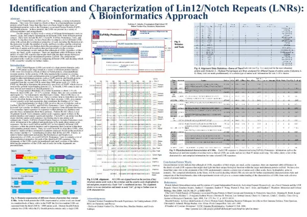
Identification and characterization of minimal ARNT-binding AINT fragments. Laura Salguero, † Carrie Partch, ‡ and Kevin Gardner ‡ † New Mexico State University, Department of Physics ‡ UT Southwestern Medical Center, Departments of Biochemistry and Pharmacology. Overview.

E N D
Identification and characterization of minimal ARNT-binding AINT fragments Laura Salguero,† Carrie Partch,‡ and Kevin Gardner‡ †New Mexico State University, Department of Physics ‡UT Southwestern Medical Center, Departments of Biochemistry and Pharmacology
Overview The HIF (Hypoxia Inducible Factor) transcription factor is composed of two proteins: HIF- and HIF- (a.k.a. ARNT ARyl hydrocarbon Nuclear Translocator). These two proteins bind each other in response to specific signals from the cell that it has encountered low oxygen levels; most of these interactions occur via protein-protein interaction domains called PAS (Per-ARNT-Sim) domains in each protein. Previously, NMR spectroscopy and X-ray crystallography was used to solve the structures of a heterodimeric complex of PAS domains from both HIF-2 and ARNT: This structure has provided insights both into the nature of the interaction and information about how it might be artificially controlled. Controlling HIF signaling is particularly important because oxygen can only diffuse ca. 100m in living tissue, causing solid tumors to send out a variety of biochemical signals that trigger new blood vessel formation and other responses that let them adapt to low oxygen levels. This project was focused on studying the next step in hypoxia signaling: how the HIF- heterodimer recruits a coactivator protein, AINT (ARNT INTeracting protein). This is of particular interest to us in that initial data suggests that AINT interacts directly with the ARNT PAS-B domain, implicating a new type of protein-protein interaction that PAS domains might be able to participate in. This also firmly indicates that these PAS domains can actually simultaneously bind two different protein partners at the same time. The structure of such a novel complex is unknown at this point, and is of intense interest.
HIF-1 complex is an oxygen-sensitive transcriptional switch growth of solid tumors • promotion of angiogenesis, or formation of new blood vessels • activation of glucose transport and glycolysis • induction of erythropoeisis, the formation of new red blood cells
bHLH PAS-A PAS-B Card, P.B. et al (2005) J Mol Biol Tom Scheuermann (2008) • Transcription factors must recruit diverse co-activator proteins to initiate transcription • Modification of local chromatin structure • Recruitment of transcriptional machinery • AINT is a co-activator that interacts with nuclear Histone AcetylTransferases (HAT), that modify histones to stimulate transcription. (Gangisetty, O et al (2004) Oncogene) • AINT binds to ARNT PAS-B and over expression of it stimulates the transcriptional response to hypoxia by HIF- and ARNT. (Sadek, C et al (2000) Mech. of Dev.) Co-activator recruitment by ARNT ARNT domain structure Hypothesis ARNT Since the sheet of ARNT PAS-B is involved in binding to HIF-as shown, the helices on ARNT PAS-B must be binding to AINT. AINT binding? Hif-2
Goals of Project 1.) Identify minimal region of AINT that interacts with ARNT PAS-B • express GST-tagged AINT fragments in E.coli and test interaction with His-tagged ARNT PAS-B by pulldown assay • Map interaction site on 15N ARNT PAS-B by 2D protein NMR The ARNT binding domain of AINT is the C-terminal coiled-coil domain (Sadek, C et al (2000) Mech of Dev). Since coiled-coil structures are highly elongated, it is desirable to identify the smallest segment representing the ARNT binding sequence while keeping coiled-coil structure. Sec2p coiled-coil (150 res.) Sec4p (169 res.) Dong, G. et al (2007) Mol Cell 2.) Purify AINT minimal domain using HisGB1-tag and monitor the coiled-coil structure by circular dichroism spectroscopy 3.) Set up crystallization trays with an ARNT PAS-B:AINT complex for high resolution structure determination
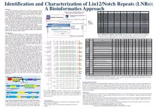
,His-ARNT PAS-B 10% Input , GST-AINT: 4 8 6 7 9 10 11 4 8 6 7 9 10 11 62 47.5 32.5 25 16.6kDa AINT ARNT PAS-B GST His Ni2+ resin Identification of Minimal AINT Fragment A pulldown assay is a technique to assess protein-protein interactions. Soluble E.coli extract with GST-AINT fragments was incubated with His-ARNT PAS-B and Ni2+ resin. Protein binding was assessed after pelleting the resin, washing, and resolving the mix by denaturing gel electrophoresis. Conclusion: AINT 10 is smallest fragment (40 amino acids) tested with binding to ARNT PAS-B and the C-terminal 20 residues are essential for binding.
IFT Eluate , Imidazole 97 66 45 30 20.1 14.4 kDa Time (m):0 2 5 10 20 60 180 14.4 kDa Dimer Monomer Purification of AINT 10 • His-GB1 AINT 10 was expressed in E.coli BL21(DE3) cells; soluble extract was run over a Ni2+ column. The His-GB1 tag attaches to the Ni2+ resin and debris is washed away in the flow through. Protein was eluted with a gradient of imidazole. • His-TEV (Tobacco Etch Virus) protease was added to cleave the His-GB1 tag; protease and tag were removed by Ni2+ column. • Each monomer of AINT 10 has only one cysteine suggesting that it forms a native disulfide bond as a coiled-coil dimer, therefore CuSO4 was used to oxidize the cysteines and induce disulfide formation. • 1mM AINT10 was incubated with 1mM CuSO4 at room temperature for three hours. The reaction was quenched with 2mM EDTA. AINT 10 - 5,473.3 g/mol EVLALQASLRKAQMQNHSLEMTLEQKTKEIDELTRICDDLISKMEKI EVLALQASLRKAQMQNHSLEMTLEQKTKEIDELTRICDDLISKMEKI NaCl Input Eluate , 97 66 45 30 20.1 14.4 kDa • Dimer was run over a Mono S cation exchange column to remove CuSO4 and further contaminates, and buffer exchanged to dilute the high salt required to elute AINT 10 from the column.
Far UV CD is a spectroscopic technique that measures the arrangement of peptide bonds in secondary structure elements like helices and strands Circular Dichroism (CD) Spectrum of AINT 10 Ref: www.photophysics.com/circulardichroism.php • Acquired in Aviv 62D Spectropolarimeter at 25C • 15M disulfide-linked AINT 10 dimer • 10mM Sodium Phosphate pH 6.5, 17mM NaCl Conclusion: AINT 10 is a parallel coiled-coil dimer
Mapping AINT 10 binding to ARNT PAS-B • 15N-1H Heteronuclear Single Quantum Coherence (HSQC) spectrum is a • ‘protein fingerprint’ where each crosspeak corresponds to a backbone amide • This titration experiment monitors the perturbation of 15N ARNT PAS-B with • the addition of unlabeled AINT 10 SS Dimer Conclusions: AINT 10 appears to bind on to the helical face of ARNT PAS-B
http://www-structmed.cimr.cam.ac.uk/Course/Basic_diffraction/Diffraction.xhthttp://www-structmed.cimr.cam.ac.uk/Course/Basic_diffraction/Diffraction.xht X-ray Crystallography • For determining protein structures X-ray crystallography can be fast and provide high resolution structural details if a protein will crystallize nicely. • Crystallography is a technique that determines crystalline structures using Bragg’s Law, n=2dsin() • X-rays are generated and focused to maximum intensity, here at 8keV, and diffracted off a crystal at multiple angles in order to see multiple planes within the crystal. • Since x-rays will have both constructive and destructive interference, the diffraction pattern resembles dots in a ring around a central position. These spots represent a spacing between atoms which is inversely proportional to the distance of the ring to the center. The closer the spots are to the center, the larger distance they represent.
Crystal Screens • All protein have different conditions at which they want to crystallize. As there is no way of knowing what these conditions may be for a novel protein, a screen is set up that tests the protein under a wide variety of precipitants, pHs and buffers. • All of our crystal screening trays, except three of the optimization trays, were set up using the Phoenix robot in the Structural Biology Lab: • QIAGEN The Classics • QIAGEN The JCSG+ Suite • QIAGEN The Protein Complex Suite • QIAGEN The PEGs Suite • Hampton Salt Rx • Hampton Index • From these screens, over 170 conditions formed crystals. Four optimization trays and one streak-seeding tray has been set up. This effort will be continued into the future.
Conclusions Acknowledgements • Found minimal domain of AINT necessary for binding ARNT • Mapped the binding on ARNT PAS-B to the -helices • Showed by CD that AINT 10 is parallel coiled-coil dimer • Crystallography of the protein complex in process Thank you to: The Gardner lab: Jason Key, Abby Nash, Matthew Evans, Kim Shahi, Amy Zhou, Qiong Wu, Abby Nash, Lisa Ko, Tom Scheuermann, Paul Card, Fernando Correa, Qun Du Diana Tomchick and the Structural Biology lab Charles Dann III Carlos Amezcua and the Biomolecular NMR Facility Quantitative & Physical Sciences Summer Training Program; NIGMS 5R25GM072832-04 Minority Access to Research Careers; GMO7667-31 This work could provide critical understanding of the transcriptional response to hypoxia and help develop chemotherapeutic intervention.