Download
1 / 47
580 likes | 1.04k Views
Blood. Objectives. List the three primary functions of blood List avg vol. of blood, viscosity, pH, % body weight Distinguish between plasma and formed elements

E N D
Objectives • List the three primary functions of blood • List avg vol. of blood, viscosity, pH, % body weight • Distinguish between plasma and formed elements • Identify substances in plasma and distinguish among the 3 categories of proteins based on their purpose (albumins, globulins, fibrinogens) • Explain the procedure for hematocrit separation • List the 3 types of formed elements • List primary purpose of erythrocyte • Describe the composition of hemogloblin • List the life span of an erythrocyte and the destruction/recycling of the components when it is aged • Define hematopoiesis and list where in the body it occurs • List dietary requirements for erythropoiesis to occur
Functions of Blood • Blood performs a number of functions dealing with: • Substance distribution • Brings: nutrients from dig. organs, O2 from lungs, hormones from endocrine glands • Take: waste (CO2, urea, uric acid, etc) • Regulation of blood levels of particular substances • Acid/Base balance of body fluids (buffer) • Vol. of blood flow to diff. areas of body • Body protection • Phagocytic wbc and antibodies • Release clotting factors
Physical Characteristics of Blood • Characteristics indicate individual health state • Average volume of blood: • 5–6 L for males; 4–5 L for females (Normovolemia) • Hypovolemia - low blood volume • Hypervolemia - high blood volume • Viscosity (thickness) - 4 - 5 (where water = 1) • Due to formed elements (RBC) • Flows 5x slower than water • The pH of blood is 7.35–7.45 • Blood accounts for approximately 8% of body weight
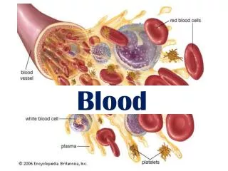
Composition of Blood • Blood is the body’s only fluid tissue (a connective tissue) • 2 major components • Liquid = plasma (55%) • Formed elements (45%) • Erythrocytes, or red blood cells (RBCs) • Leukocytes, or white blood cells (WBCs) • Platelets - fragments of megakaryocytes in marrow
Components of Whole Blood Plasma(55% of whole blood) Buffy coat:leukocyctes and platelets(<1% of whole blood) Formed elements Erythrocytes(45% of whole blood) Withdraw blood and place in tube Centrifuge 1 2 • Hematocrit • Males: 47% ± 5% • Females: 42% ± 5%
Blood Plasma • Blood plasma components: • Clear, yellowish fluid suspends formed elements • Water = 90-92% • Proteins = 6-8% • Approx. 50 diff types found, classified into categories • 3 Categories • Albumins • Thicken blood • Globulins • Serve as antibodies for immune response • Fibrinogen • Serve as clotting proteins
Blood Plasma • Additional dissolved substances (2%) • Organic nutrients – glucose, carbohydrates, amino acids • Electrolytes – sodium, potassium, calcium, chloride, bicarbonate • Nonprotein nitrogenous substances – lactic acid, urea, creatinine • Respiratory gases – oxygen and carbon dioxide
Formed Elements • Formed elements comprise 45% of blood • Erythrocytes, leukocytes, and platelets make up the formed elements • Only WBCs are complete cells • RBCs have no nuclei or organelles, and platelets are just cell fragments • Most formed elements survive in the bloodstream for only a few days • Most blood cells do not divide but are renewed by cells in bone marrow
Erythrocytes RBCs
Erythrocytes Characteristics • Biconcave disc • Folding increases surface area (30% more surface area) • Anucleate, no centrioles, no organelles • End result - no cell division • No mitochondria means they generate ATP anaerobically • Prevents consumption of O2 being transported • Filled with hemoglobin (Hb) - 97% of cell contents • Hb functions in gas transport • Most numerous of the formed elements • Females: 4.3–5.2 million cells/cubic millimeter • Males: 5.2–5.8 million cells/cubic millimeter
Erythrocytes (RBCs) Figure 17.3
Erythrocyte Function • Erythrocytes are dedicated to respiratory gas transport • Hemoglobin reversibly binds with oxygen and most oxygen in the blood is bound to hemoglobin • Composition of hemoglobin • A protein called globin • made up of two alpha and two beta chains • A heme molecule • Each heme group bears an atom of iron, which can bind to one oxygenmolecule • Each hemoglobin molecule thus can transport four molecules of oxygen
Structure of Hemoglobin Figure 17.4
Hemoglobin • Oxyhemoglobin – hemoglobin bound to oxygen • Oxygen loading takes place in the lungs • Deoxyhemoglobin – hemoglobin after oxygen diffuses into tissues (reduced Hb) • Carbaminohemoglobin – hemoglobin bound to carbon dioxide • Carbon dioxide loading takes place in the tissues
Fate and Destruction of Erythrocytes • The life span of an erythrocyte is 100–120 days • Travels about 750 miles in that time • Old erythrocytes become rigid and fragile, and their hemoglobin begins to degenerate • Dying erythrocytes are engulfed by macrophages • Heme and globin are separated • Iron is removed from the heme and salvaged for reuse • Stored as hemosiderin or ferritin in tissues • Transported in plasma by beta-globulins as transferrin
Fate and Destruction of Erythrocytes • Heme is degraded to a yellow pigment called bilirubin • Liver secretes bilirubin into the intestines as bile • Intestines metabolize bilirubin into urobilinogen • Urobilinogen leaves the body in feces, in a pigment called stercobilin • Globin is metabolized into amino acids which are then released into the circulation
Stages of Differentiation of Blood Cells Figure 17.9
Production of Erythrocytes • Hematopoiesis – blood cell formation • Occurs in the red bone marrow (myeloid tissue) • Axial skeleton and girdles • Epiphyses of the humerus and femur • Marrow contains immature erythrocytes • Hemocytoblasts give rise to ALL formed elements • Lymphoid stem cells - give rise to lymphocytes • Myeloid stem cells - give rise to all other blood cells
Regulation and Requirements for Erythropoiesis • Circulating erythrocytes – the number remains constant and reflects a balance between RBC production and destruction • Too few red blood cells leads to tissue hypoxia • Too many red blood cells causes undesirable blood viscosity • Erythropoiesis is hormonally controlled and depends on adequate supplies of iron, amino acids, and B vitamins
Hormonal Control of Erythropoiesis • Erythropoietin (EPO) release by the kidneys is triggered by: • Hypoxia due to decreased RBCs • Decreased oxygen availability • Increased tissue demand for oxygen • Enhanced erythropoiesis increases the: • RBC count in circulating blood • Oxygen carrying ability of the blood
Erythropoietin Mechanism Imbalance Start Normal blood oxygen levels Stimulus: Hypoxia due to decreased RBC count, decreased availability of O2 to blood, or increased tissue demands for O2 Imbalance Increases O2-carrying ability of blood Reduces O2 levels in blood Erythropoietin stimulates red bone marrow Kidney (and liver to a smaller extent) releases erythropoietin Enhanced erythropoiesis increases RBC count Figure 17.6
Dietary Requirements of Erythropoiesis • Erythropoiesis requires: • Proteins, lipids, and carbohydrates • Iron, vitamin B12, and folic acid • The body stores iron in Hb (65%), the liver, spleen, and bone marrow • Intracellular iron is stored in protein-iron complexes such as ferritin and hemosiderin • Circulating iron is loosely bound to the transport protein transferrin
Erythrocyte Disorders • Polycythemia • Abnormal excess of erythrocytes • Increases viscosity, decreases flow rate of blood • Leads to rise in BP • If untreated can cause blood clots (thrombosis) and hemorrhage • Anemia • Anemia • Reduction in # of RBCs or in amt of Hg per unit of blood • Several types • It is a symptom rather than a disease itself • Blood oxygen levels cannot support normal metabolism • Signs/symptoms include fatigue, paleness, shortness of breath, and chills
Anemia: Insufficient Erythrocytes • Hemorrhagic anemia • Result of acute or chronic loss of blood • Hemolytic anemia • Prematurely ruptured erythrocytes • Aplastic anemia • Destruction or inhibition of red bone marrow • aplastic anemia
Anemia: Decreased Hemoglobin Content • Iron-deficiency anemia results from: • A secondary result of hemorrhagic anemia • Inadequate intake of iron-containing foods • Impaired iron absorption • Pernicious anemia results from: • Deficiency of vitamin B12 • Lack of intrinsic factor needed for absorption of B12 • Treatment is intramuscular injection of B12
Anemia: Abnormal Hemoglobin • Thalassemias • Absent or faulty globin chain in hemoglobin • Erythrocytes are thin, delicate, and deficient in hemoglobin • documentary • Sickle-cell anemia • Results from a defective gene • Codes for an abnormal hemoglobin called hemoglobinS (HbS) • This defect causes RBCs to become sickle-shaped in low oxygen situations
Leukocyte Objectives • Differentiate between leukocytosis, leukopenia and leukemia • Differentiate between two main categories of leukocytes • Name the 3 different types of lymphocytes listing each purpose of the specific cell type • Be able to discuss differences between lymphocytes and monocytes in terms of quantity, function and structure • List features of the granular cells that are different that agranular cells ( ability to be stained, life span, nuclei) • Name the 3 types of granular cells- distinguish between them in terms of %, function, life span • Explain PMN/poly abbreviation for neutrophils • Define leukemia,origin of different types, treatment • Define platelets and explain function
Leukocytes (WBCs) • Leukocytes, the only blood components that are complete cells: • 4,800 - 10,000/cubic millimeter • Leukocytosis • WBC count over 11,000/mm3 • Normal response to bacterial or viral invasion • Leukopenia • A decrease in WBC count below 4,800/mm3 • Leukemia - a cancer of WBC
Leukocytes • Purpose • Mobile units of body’s defense system • “Seek and Destroy” Functions • Destroy invading microorganisms • Destroy abnormal cells (ie: cancer) • Clean up cellular debris (phagocytosis)
Leukocyte Type • Two main categories • Agranular • Lymphocytes • Monocytes • Granular • Neutrophils • Eosinophils • Basophils
5 - Types of WBC’s Granulocytes Agranulocytes Each WBC has a specific function
Agranulocytes: Lymphocytes • Account for 25-30% or more of WBCs and: • Absence of granules in cytoplasm when stained (however do contain lysosomes) • Most important cells of the immune system • There are three types of lymphocytes: • T cells • B cells • Natural killer cells
Agranulocytes: Lymphocytes • T cells - attack foreign cells directly, helps restore immune system after infection; (cell- mediated immunity) • B cells - produce antibodies that bind to pathogens to enable their destruction (humoral immunity) • Natural killer cells – kills cells of the body that are displaying a signal to them (due to virus/cancer infection)
Agranuloctes: Monocytes • Monocytes account for 3–7% of leukocytes • They are the largest leukocytes • U- or kidney-shaped nuclei • They leave the circulation, enter tissue, and differentiate into macrophages • Clean up dead cell debris and attack microorganisms
Agranuloctes: Monocytes Animation Microscope
Granulocytes • Granulocytes – neutrophils, eosinophils, and basophils • Contain cytoplasmic granules that stain specifically (acidic, basic, or both) with Wright’s stain • Are larger and usually shorter-lived than RBCs • Have lobed nuclei • Are all phagocytic cells
Granulocytes: Neutrophils(Polymorphonuclear leukocytes) • Account for 60-70% of total WBC’s • Life span short (~ 5 days) • Function • Neutrophils are our body’s bacteria slayers • AKA “polys” or PMN’s because multi-lobed nucleus can appear has multiple nuclei(polymorphonuclear) • Present in large amounts of pus
Granulocytes: Eosinophils • Eosinophils account for 1–4% of WBCs • Life span short (~10 days) • Function • Lead the body’s counterattack against parasitic infections • Lessen the severity of allergies by phagocytizing immune complexes (ending allergic reactions)
Granulocytes: Basophils • Rare:Account for 0.5-1% of all WBCs • Life span short (~few hours – few days) • Function • Release histamine for inflammatory response • Histamine – inflammatory chemical that acts as a vasodilator and attracts other WBCs (antihistamines counter this effect)
Formation of Leukocytes • All leukocytes originate from hemocytoblasts • The mother of all blood stem cells • Hemocytoblasts differentiate into myeloid stem cells and lymphoid stem cells • Myeloid stem cells become myeloblasts or monoblasts • Granulocytes form from myeloblasts • Monoblasts enlarge and form monocytes • Lymphoid stem cells become lymphoblasts • Lymphoblasts develop into lymphocytes
Formation of Leukocytes Figure 17.11
Leukocytes Disorders: Leukemias • Leukemia refers to cancerous conditions involving white blood cells • Leukemias are named according to the abnormal white blood cells involved • Myelocytic leukemia – involves myeloblasts • Lymphocytic leukemia – involves lymphocytes • Acute leukemia involves blast-type cells and primarily affects children • Chronic leukemia is more prevalent in older people
Leukemia • Immature white blood cells are found in the bloodstream in all leukemias • Bone marrow becomes totally occupied with cancerous leukocytes • Severe anemia ensues due to excess production of WBC’s • The white blood cells produced, though numerous, are not functional • Death is caused by internal hemorrhage and overwhelming infections • Treatments include irradiation, antileukemic drugs, and bone marrow transplants
Platelets • Platelets are fragments of megakaryocytes • Platelets function in the clotting mechanism by forming a temporary plug that helps seal breaks in blood vessels


