Understanding Hormones and Endocrine Pathways in Human Physiology
280 likes | 402 Views
This chapter explores the intricate workings of hormones and the endocrine system, detailing various hormonal pathways that regulate vital bodily functions. It illustrates mechanisms such as low blood glucose response, the roles of the hypothalamus and pituitary glands, and the effects of hormones like glucagon, insulin, and oxytocin. Through figures and summaries, it provides insight into endocrine cell signaling, feedback regulation, and the impact of hormones on growth, metabolism, and homeostasis. Key examples underscore the complex interactions that maintain physiological balance.

Understanding Hormones and Endocrine Pathways in Human Physiology
E N D
Presentation Transcript
Chapter 45 Hormones and theEndocrine System
Figure 45.1 An anise swallowtail butterfly emerging from its chrysalis
Pathway Example Pathway Example Example Pathway Low blood glucose Hypothalamic neurohormone released in response to neural and hormonal signals Stimulus Stimulus Suckling Stimulus Receptor protein Sensory neuron Sensory neuron Pancreas secretes glucagon ( ) Hypothalamus/ posterior pituitary Hypothalamus Endocrine cell Neurosecretory cell Blood vessel Neurosecretory cell Hypothalamus secretes prolactin- releasing hormone ( ) Posterior pituitary secretes oxytocin ( ) Blood vessel Blood vessel Target effectors Liver Anterior pituitary secretes prolactin ( ) Smooth muscle in breast Target effectors Glycogenbreakdown,glucose releaseinto blood Response Endocrine cell Blood vessel (a) Simple endocrine pathway Milk release Response (b) Simple neurohormone pathway Target effectors Mammary glands Milk production Response (c) Simple neuroendocrine pathway Figure 45.2 Basic patterns of simple hormonal control pathways
SECRETORY CELL SECRETORY CELL Hormone molecule Hormone molecule VIA BLOOD VIA BLOOD Signal receptor TARGET CELL TARGET CELL Signal transduction pathway Signal receptor OR Cytoplasmic response DNA Signal transduction and response mRNA DNA NUCLEUS Nuclear response Synthesis of specific proteins NUCLEUS (b) Receptor in cell nucleus (a) Receptor in plasma membrane Figure 45.3 Mechanisms of hormonal signaling: a review
Different receptors different cell responses Epinephrine Epinephrine Epinephrine b receptor a receptor b receptor Glycogendeposits Vessel dilates Vessel constricts Glycogen breaks down and glucose is released from cell (a) Intestinal blood vessel Skeletal muscle blood vessel (b) (c) Liver cell Different intracellular proteins different cell responses Figure 45.4 One chemical signal, different effects
Figure 45.5 Activated platelets aggregating, a process regulated in part by prostaglandins
Table 45.1 Major Human Endocrine Glands and Some of Their Hormones
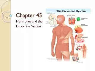
Hypothalamus Pineal gland Pituitary gland Thyroid gland Parathyroid glands Adrenal glands Pancreas Ovary (female) Testis (male) Figure 45.6 Human endocrine glands surveyed in this chapter
Hypothalamus Neurosecretory cells of the hypothalamus Axon Posterior pituitary Anterior pituitary ADH HORMONE Oxytocin Mammary glands, uterine muscles TARGET Kidney tubules Figure 45.7 Production and release of posterior pituitary hormones
Tropic Effects Only FSH, follicle-stimulating hormone LH, luteinizing hormone TSH, thyroid-stimulating hormone ACTH, adrenocorticotropic hormone Nontropic Effects Only Prolactin MSH, melanocyte-stimulating hormone Endorphin Nontropic and Tropic Effects Growth hormone Neurosecretory cells of the hypothalamus Portal vessels Hypothalamic releasing hormones (red dots) Endocrine cells of the anterior pituitary Pituitary hormones(blue dots) Endorphin FSH and LH TSH MSH HORMONE ACTH Prolactin Growth hormone Bones Mammary glands Testes or ovaries Melanocytes Liver Pain receptors in the brain Thyroid Adrenal cortex TARGET Figure 45.8 Production and release of anterior pituitary hormones
Group Work • Ever play pin the tail on the donkey? • Ever play pin the hormone on the gland? • Well we are going to make this game and send it to Hasbro. • You need to: • Draw a hermaphrodite on large paper • Draw and cut out hormone pieces • The pieces should relate to their function
Hypothalamus Anterior pituitary TSH Thyroid T3 T4 + Figure 45.9 Feedback regulation of T3 and T4 secretion from the thyroid gland TRH
Figure 45.10 Graves’ disease, the most common form of hyperthyroidism in humans
Calcitonin Thyroid gland releases calcitonin. Reduces Ca2+ uptake in kidneys Stimulates Ca2+ deposition in bones Blood Ca2+ level declines to set point STIMULUS: Rising blood Ca2+ level Homeostasis: Blood Ca2+ level (about 10 mg/100 mL) STIMULUS: Falling blood Ca2+ level Blood Ca2+ level rises to set point Stimulates Ca2+ release from bones Parathyroid gland PTH Increases Ca2+ uptake in intestines Stimulates Ca2+ uptake in kidneys Active vitamin D Figure 45.11 Hormonal control of calcium homeostasis in mammals
Body cells take up more glucose. Insulin Beta cells of pancreas are stimulated to release insulin into the blood. Liver takes up glucose and stores it as glycogen. STIMULUS: Rising blood glucose level (for instance, after eating a carbohydrate- rich meal) Blood glucose level declines to set point; stimulus for insulin release diminishes. Homeostasis: Blood glucose level (about 90 mg/100 mL) Blood glucose level rises to set point; stimulus for glucagon release diminishes. STIMULUS: Dropping blood glucose level (for instance, after skipping a meal) Alpha cells of pancreas are stimulated to release glucagon into the blood. Liver breaks down glycogen and releases glucose into blood. Glucagon Figure 45.12 Maintenance of glucose homeostasis by insulin and glucagon
Stress Nerve signals Hypothalamus Spinal cord (cross section) Releasing hormone Nerve cell Anterior pituitary Blood vessel Adrenal medulla secretes epinephrine and norepinephrine. Nerve cell Adrenal cortex secretes mineralocorticoids and glucocorticoids. ACTH Adrenal gland Kidney (a) Short-term stress response (b) Long-term stress response Effects of glucocorticoids: Effects of mineralocorticoids: Effects of epinephrine and norepinephrine: 1. Glycogen broken down to glucose; increased blood glucose Proteins and fats broken down and converted to glucose, leading to increased blood glucose 1. Retention of sodium ions and water by kidneys 1. 2. Increased blood pressure 3. Increased breathing rate 4. Increased metabolic rate 2. Increased blood volume and blood pressure 5. Change in blood flow patterns, leading to increased alertness and decreased digestive and kidney activity 2. Immune system may be suppressed Figure 45.13 Stress and the adrenal gland
Figure 45.14 Male breast enlargement due to anabolic steroids
Brain Neurosecretory cells Brainhormone (BH) Corpus cardiacum Corpus allatum Prothoracicgland Ecdysone EARLYLARVA LATERLARVA PUPA ADULT Figure 45.15 Hormonal regulation of insect development (layer 1)
Brain Neurosecretory cells Brainhormone (BH) Corpus cardiacum Corpus allatum Prothoracicgland Ecdysone EARLYLARVA LATERLARVA PUPA ADULT Figure 45.15 Hormonal regulation of insect development (layer 2)
Brain Neurosecretory cells Brainhormone (BH) Corpus cardiacum Corpus allatum LowJH Prothoracicgland Ecdysone Juvenilehormone(JH) EARLYLARVA LATERLARVA PUPA ADULT Figure 45.15 Hormonal regulation of insect development (layer 3)