Download
1 / 35
350 likes | 546 Views
Objectives :. Know the stages of cellular division- mitosis and meiosis. Understand the process of cytokinesis and how it differs in plants and animals. Know the differences in mitosis and meiosis. Understand the important process of mitosis and meiosis. Cellular Division.

E N D
Objectives : Know the stages of cellular division- mitosis and meiosis. Understand the process of cytokinesis and how it differs in plants and animals. Know the differences in mitosis and meiosis. Understand the important process of mitosis and meiosis.
Cellular Division • Cell division – necessary for the formation of multicellular organisms • from fertilized egg. • Cell division – 2 processes: • division of nucleus ( 2 types – mitosis and meiosis). • cytokinesis(division of the cytoplasm and other nonnuclear compounds of the cell to form 2 daughter cells).
CHROMOSOME TRANSMISSION AND CELLULAR REPRODUCTION – MITOSIS AND MEIOSIS • In all organisms, transmission of genetic material by component called Chromosome must occur – in order to propagate parental genetic material into offsprings. • Its depending on the type of organisms. • In prokaryotes, chromosome undergoes mitosis. • In eukaryotes, transmission of genetic material from one generation of cells to the next involves : • (a) mitosis – for somatic cells. • (b) meiosis – for gametes and spores.
Chromosome – threadlike structures consisting mainly of a complex of DNA (genetic material), RNA and proteins. Chromosome have key morphological features such as centromeres (join the two sister chromatic/two identical copies of parental chromosomes), kinetochore (protein structure at the centromers – functions in chromosome movement during cell division) and telomers (end of chromosomes). Diagram 1: The structure of a highly condensed, replicated chromosome
Eukaryotic Chromosomes • Eukaryotic chromosomes are inherited as sets: • Majority of eukaryotic species are diploid, thus all somatic cells • have two sets of chromosomes. • - Pairs of the same chromosomes are called homologues. • Homologues chromosomes are very similar, but may contain • different alleles of the same gene. • - Sex chromosomes are not homologues. • Genes on homologues chromosomes have the same location, or loci. • Involved in 2 main activities: • The transmission of genetic information from cell to cell and from generation to generation. • The proper expression of this information to control cellular function and development.
The Cell Cycle • Organisms to grow – must take place 3 events: • 1. Cell mass must increase. • 2. There must be a duplication of the genetic material. • 3. A division process must occur – so that each daughter cell • receives an equal and identical complement of the genetic material • (to ensure perpetuation of cell line). • Eukaryotic cells – undergo a cell cycle that consists of several distinct phases: • a. G1 and G2 (gap phases), S (synthesis) and M (mitosis). • b. G1, G2 and S phases are collectively called interphase. • c. Some cells remain in G0 phase (just prior to S phase) for extended periods of time, thus arresting cell division.
Diagram 2:cell cycle • The cell cycle is composed of interphase and mitosis. Interphase includes S phase, during which DNA is synthesized, and two gap phases (G1 and G2). G0 is a point in the G1 phase where cells withdraw from the cell cycle and enter a nondividing but metabolically active state (proteins and cytoplasmic organelles are synthesized). Thus, a cell grows (G1), continues to grow as it duplicates its chromosomes (S), grows more and prepares for mitosis (G2), and divides (M).
Preparation of cells division begins in the G1 phase. Molecular • changes accumulate in the cell, allowing it to pass through a • restriction point and into S phase. • In S phase – chromosomes are replicated, forming the sister • chromatids. Sister chromatids are linked together and are • considered a single chromosome. At this point, the cell technically • has twice as many chromosomes (46 pairs of sister chromatids) and into the G2 phase to prepare for mitosis phase. • In M phase (mitosis) – the cell distributes the replicated chromosomes so that each of the new daughter cells has an exact complement of chromosomes that were found in the original cell. • M phase – signals the actual division of the cell.
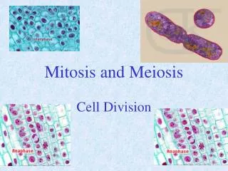
A. Mitosis (somatic cells) • Mitosis – proceeds through 5 phases; prophase, prometaphase, metaphase, anaphase, telophase. (cytokinesis follows telophase) • Mitosis leads to production of two cells, each with the same number of chromosomes as the parent cell. • Eg: • Parent diploid cell (2n) Daughter cell (2n) Daughter cell (2n) Daughter cell (n) Parent haploid cell (n) Daughter cell (n) • Genetic material is partitioned to daughter cells during nuclear division (karyokinesis). • Cytoplasmicdivision (cytokinesis) follows.
Prophase • - nuclear membrane dissociates and the chromosomes condense. • mitotic spindle apparatus forms, originating at the centromers • 3 types mitotic spindle: • Aster microtubules emanate away from the chromosomes and are important in the positioning of the spindle fibers in the cell. • Polar microtubules project toward the chromosomes and are involved in the separation of the two poles. • Kinetochore microtubules attach to the kinetochore, which in turn is connected to the centromer of the chromosome. • - centriol move apart. • - end of prophase, chromosome in replicated form. • - 2 sister chromatids held together at centromer. • - nucleolus dispersed.
Prometaphase • Interaction of the spindle fibers with the chromosome occurs. • Once a kinetochore microtubule comes in contact with the kinetochore, it is captured and no longer moves. • Kinetochoremicrotubules connect to the kinetochorefrom both poles of the cell, and gently tug the chromosomes back and forth during metaphase. • Nuclear membrane break down.
Metaphase • occurs when the chromosomes align along a central plane called the metaphase • plate. • centriolreached at opposite poles. See diagram 5. • Anaphase • - sorting of chromosomes occurs during anaphase,whenthe connection between the sister chromatidsbreaks. • at this point, each of the chromatids is linked to only one of the poles. • each chromatid is now considered to be an independent chromosome. • the chromosomes now begin to migrate to their respective poles of the cell. • See diagram 6. • Telophase • when the cell reach opposite sides of the cell, they begin to condense (this marks the start of telophase). • nuclear membrane then reforms around the chromosomes. See diagram 7.
Cytokinesis- following telophase, cell proceeds into cytokinesis or cytoplasmic division. • - in animal cells – involved the use of a cleavage furrow. • - in plant cells – involved the use of a cell plate. • The end result of mitosis and cytokinesis = 2 daughter cells that are genetically identical. • Small variations are possible due to mutation. • Interphase - alternate with mitosis. • - cell grows occur and preparation for the next mitosis process. • - 3 steps: DNA synthesis (S) and 2 steps of cell growth (G1 and G2).
B. Meiosis (gamete cells) • Sexual Reproduction: Involve Meiosis (cell division). • The purpose of sexual reproduction – to produce gametes that will combine by the process of fertilisation to produce new individual (zygote). • Gametes – are haploid (n) cells that contain a single set of chromosomes. Gametes are produced from diploid (2n) cells by the process of meiosis.
A process in sexual reproduction through which the chromosome number of diploid (2n) germ cells is reduced to half (n) in formation of mature reproductive cells or gametes. • Similar to mitosis in many aspect, except that it involves • 2 consecutive cell divisions within an intervening interphase. • Meiosis I – the homologous chromosomes come together in pairs and duplicate. Each pair of homologous chromosome enter different daughter cell. • Daughter cell = ½ parent chromosome. • Meiosis II – similar to mitosis, sister chromatids (of each chromosome) separated, • and enter into different cells, making a total of four separate mature sex cells each capable of fertilisation. • Through fertilisation the diploid (2n) chromosome number is restored. An organism characteristic of a species may then be developed largely by mitotic cell division. • Therefore, meiosis – produced 4 daughter cells (n) that vary in their genetic composition. Mitosis produced 2 daughter cells (2n) with identical genetic composition from the parental cells.
Meiosis leads to production of four daughter cells, • each with half the number of chromosomes of the parent cell. • Takes place in germ cells and spores. • Meiosis reduces the amount of genetic material by one-half to produce haploid gametes or spores containing one member of each homologous pair of chromosomes. • Meiosis is divided to two major cycles: Meiosis I & Meiosis II. • Meiosis I is a reductionaldivision. • Meiosis II is an equational division (resembles mitosis). • DNA synthesis/replication occurs during interphase before the beginning of meiosis I but does not occur again before meiosis II.
Meiosis I: Prophase I i. Leptonema - chromosomes start to condense, forming threadlike structures. ii. Zygotene/zygonema - the homologous chromosomes recognise each other by a process known as synapsis. - chromosomes align along their entire lengths. - at this point, the pairs of homologous chromosomes are called bivalents (4 sister chromatids in a bivalent). - synaptonemal complex forms. iii. Pachytene/Pachynema - just prior to this third stage, process of crossing over occurs between non-sister chromatidsin the bivalent. - the site of crossing over – chiasma. iv. Diplotene/Diplonema - the synaptonemal complex has disappeared. The individual chromatids are usually visible at this point, and the structure called a tetrad (4 chromatids). v. Diakinesis - the synaptonemal complex has completely disappeared.
Metaphase I - the chromosomes align along a central line in the cell in the same manner as mitosis, except: (i) the bivalent chromosomes are aligned in a double row (mitosis = single row) (ii) the kinetochoremicrotubules link one of the homologous chromosomes to one of the poles, while a second set of kinetochore microtubules link the other homologous chromosome to the other pole. - each bivalent pair may align in one of two configurations. - the number of different, random alignments for a species is equal to 2n, where n equals the chromosome number. Anaphase I - the homologous chromosomes separate from each other and begin to migrate to opposite poles. - the sister chromatids remain connected during Meiosis I.
Telophase I • - the chromosomes reach the opposite poles of the cell and begin to decondense. • - nuclear membrane then reforms around chromosomes. • Cytokinesis • Follows telophase I, cytokinesis occurs and the cell proceeds directly to Meiosis II (sequence of events is identical to mitosis, except that two cells are now dividing). • Cytokinesis follows Meiosis II.
Diagram 10 : Photographs showing the stages of meiosis in the plant Lilium regale.
Importance of Mitosis and Meiosis • Mitosis is vital for: • (a) asexual reproduction – produces offsprings with identical number of chromosomes with parent. (b) producing new cells for growth (c) producing new cells for tissue regeneration • Meiosis is vital for: (a) sexual reproduction - produces gametes carrying different alleles of paternal and maternal genetic information. (b) producing genetic variation among offsprings - through sexual reproduction, offspring inherit new combinations of alleles, which leads to variations in traits This variation in traits is the basis for evolutionary change. - segregation and independent assortment of gene (Mendel) – (anaphase 1). - maternal and paternal chromosome choose patners randomly from the crossing over process (pachynema - prophase 1). - paternal and maternal genes (on each chromosome: diploid), distributed into gamete via segregation, assortment and crossing over processes. Therefore, it is impossible to have gamete with the same genome. (c) Maintain the amount of chromosome in organism. - diploid – haploid gamete - combination of gamete – zygote (diploid)
Further information and understanding, visit this website: http://www.vcbio.science.ru.nl/en/virtuallessons/meiostage/ http://www.wikipedia.com
Students should be able: • Know the stages of cell cycle and recognize the diagrams of mitosis and meiosis associated with the process. • Understand the process involve in each stage of mitosis and meiosis. • Understand the process of cytokinesis. • Know the differencess between the two process and their importance in cell cycle.