Download
1 / 20
280 likes | 1.02k Views
Cell Division, Mitosis, and Meiosis.

E N D
Cell Division, Mitosis, and Meiosis Cell Division Functions in Reproduction, Growth, and Repair Cell division involves the distribution of identical genetic material, DNA, to two daughters cells. What is most remarkable is the fidelity with which the DNA is passed along, without dilution or error, from one generation to the next.
Core Concepts: • All Organisms Consist of Cells and Arise from Preexisting Cells • Mitosis is the process by which new cells are generated. • Meiosis is the process by which gametes are generated for reproduction. • The Cell Cycle Represents All Phases in the Life of a Cell • DNA replication (S phase) must precede mitosis, so that all daughter cells receive the same complement of chromosomes as the parent cell. • The gap phases separate mitosis from S phase. This is the time when molecular signals mediate the switch in cellular activity. • Mitosis involves the separation of copied chromosomes into separate cells • Unregulated Cell Division Can Lead to Cancer • Cell-cycle checkpoints normally ensure that DNA replication and mitosis occur only when conditions are favorable and the process is working correctly. • Mutations in genes that encode cell-cycle proteins can lead to unregulated growth, resulting in tumor formation and ultimately invasion of cancerous cells to other organs.
Core Concepts… • In order to better understand the concept of cell division and genetics, some basic definitions are in order: • gene - basic unit of heredity; codes for a specific trait • locus - the specific location of a gene on a chromosome (locus - plural loci) • genome - the total hereditary endowment of DNA of a cell or organism • somatic cell - all body cells except reproductive cells • gamete - reproductive cells (i.e. sperm & eggs) • chromosome - elongate cellular structure composed of DNA and protein - they are the vehicles which carry DNA in cells • diploid (2n) - cellular condition where each chromosome type is represented by two homologous chromosomes • haploid (n) - cellular condition where each chromosome type is represented by only one chromosome • homologous chromosome - chromosome of the same size and shape which carry the same type of genes • chromatid - one of two duplicated chromosomes connected at the centromere • centromere - region of chromosome where microtubules attach during mitosis and meiosis
Chromosome structure • composed of DNA and protein (histones) all tightly wrapped up in one package • duplicated chromosomes are connected by a centromere
Example - an organism is 2n = 4. Chromosomes 1 & 2 are homologous chromosomes • Chromosomes 3 & 4 are homologous chromosomes • Chromosomes 1 & 3 came from the mother • Chromosomes 2 & 4 came from the father
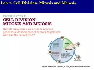
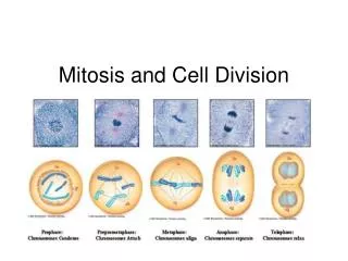
Mitosis in a Nutshell • The stages of the cell cycle can be broken down into six stages: • Interphase, Prophase, Metaphase, Anaphase, Telophase • Interphase is the "resting" or non-mitotic portion of the cell cycle. • It is comprised of G1, S, and G2 stages of the cell cycle. • DNA is replicated during the S phase of Interphase
Prophase • Prophase - the first stage of mitosis. The chromosomes condense and become visible • The centrioles form and move toward opposite ends of the cell ("the poles") • The nuclear membrane dissolves • The mitotic spindle forms (from the centrioles in animal cells) • Spindle fibers from each centriole attach to each sister chromatid at the kinetochore • Compare Prophase to the Prophase I and to the Prophase II stages of mitosis.
Metaphase • Metaphase The Centrioles complete their migration to the poles • The chromosomes line up in the middle of the cell ("the equator") • Compare Metaphase to the Metaphase I and to the Metaphase II stages of mitosis.
Anaphase • Spindles attached to kinetochores begin to shorten. • This exerts a force on the sister chromatids that pulls them apart. • Spindle fibers continue to shorten, pulling chromatids to opposite poles. • This ensures that each daughter cell gets identical sets of chromosomes • Compare Anaphase to the Anaphase I and to the Anaphase II stages of mitosis.
Telophase • The chromosomes decondense • The nuclear envelope forms • Cytokinesis reaches completion, creating two daughter cells
Cytokinesis Divides the Cytoplasm • In animal cells, cytokinesis occurs by a process known as cleavage First, a cleavage furrow appears • cleavage furrow = shallow groove near the location of the old metaphase plate • A contractile ring of actin microfilaments in association with myosin, a protein • Actin and myosin are also involved in muscle contraction and other movement functions • The contraction of a the dividing cell's ring of microfilaments is like the pulling of drawstrings • The cell is pinched in two • Cytokinesis in plant cells is different because plant cells have cell walls. • There is no cleavage furrow • During telophase, vesicles from the Golgi apparatus move along microtubules to the middle of the cell (where the cell plate was) and coalesce, producing the cell plate • Cell-wall construction materials are carried in the vesicles and are continually deposited until a complete cell wall forms between the two daughter cells
Chromosome Separation Is the Key Event of Mitosis • Mitotic spindle fibers are the railroad tracks for chromosome movement. • Spindle fibers are made of microtubules. • Microtubules are lengthened and shortened by the addition and loss of tubulin subunits. • Mitotic spindle shortening during anaphase is a result of the loss of tubulin subunits. • A kinetochore motor is the engine that drives chromosome movement. • Multiple studies have shown that the kinetochore contains motor proteins that can �walk� along the spindle fiber during anaphase. • These proteins presumably remove tubulin subunits, shortening spindle fibers and facilitating the chromosome movement.
Regulation of the Cell Cycle • The cell cycle is controlled by a cyclically operating set of reaction sequences that both trigger and coordinate key events in the cell cycle The cell-cycle control system is driven by a built-in clock that can be adjusted by external stimuli (chemical messages) • Checkpoint - a critical control point in the cell cycle where stop and go-ahead signals can regulate the cell cycle • Animal cells have built-in stop signals that halt the cell cycles and checkpoints until overridden by go-ahead signals. • Three Major checkpoints are found in the G1, G2, and M phases of the cell cycle • The G1 checkpoint - the Restriction Point • The G1 checkpoint ensures that the cell is large enough to divide, and that enough nutrients are available to support the resulting daughter cells. • If a cell receives a go-ahead signal at the G1 checkpoint, it will usually continue with the cell cycle • If the cell does not receive the go-ahead signal, it will exit the cell cycle and switch to a non-dividing state called G0 • Actually, most cells in the human body are in the G0 phase • The G2 checkpoint ensures that DNA replication in S phase has been completed successfully. • The metaphase checkpoint ensures that all of the chromosomes are attached to the mitotic spindle by a kinetochore.
Cyclins and Cyclin-Dependent Kinases - The Cell-Cycle Clock Rhythmic fluctuations in the abundance and activity of cell-cycle control molecules pace the events of the cell cycle. • Kinase - a protein which activates or deactivates another protein by phosphorylating them. • Kinases give the go-ahead signals at the G1 and G2 checkpoints • The kinases that drive these checkpoints must themselves be activated • The activating molecule is a cyclin, a protein that derives its name from its cyclically fluctuating concentration in the cell • Because of this requirement, these kinases are called cyclin-dependent kinases, or Cdk's
MPF - Maturation Promoting Factor (M-phase promoting factor)Cyclins accumulate during the G1, S, and G2 phases of the cell cycle • By the G2 checkpoint (the red bar in the figure), enough cyclin is available to form MPF complexes (aggregations of Cdk and cyclin) which initiate mitosis • MPF apparently functions by phosphorylating key proteins in the mitotic sequence • Later in mitosis, MPF switches itself off by initiating a process which leads to the destruction of cyclin • Cdk, the non-cyclin part of MPF, persists in the cell as an inactive form until it associates with new cyclin molecules synthesized during interphase of the next round of the cell cycle
PDGF - Platelet-Derived Growth Factors - An Example of an External Signal for Cell Division PDGF is required for the division of fibroblasts which are essential in wound healing • When injury occurs, platelets (blood cells important in blood clotting) release PDGF • Fibroblasts are a connective tissue cells which possess PDGF receptors on their plasma membranes • The binding of PDGF activates a signal-transduction pathway that leads to a proliferation of fibroblasts and a healing of the wound • Density Dependent Inhibition Cells grown in culture will rapidly divide until a single layer of cells is spread over the area of the petri dish, after which they will stop dividing • If cells are removed, those bordering the open space will begin dividing again and continue to do so until the gap is filled - this is known as contact inhibition • Apparently, when a cell population reaches a certain density, the amount of required growth factors and nutrients available to each cell becomes insufficient to allow continued cell growth