Download
1 / 6
80 likes | 150 Views
A 71-year-old female presented with pelvic mass and further diagnosed with myeloid sarcoma. Learn about imaging features, diagnosis, and management options in this rare gynaecological case.

E N D
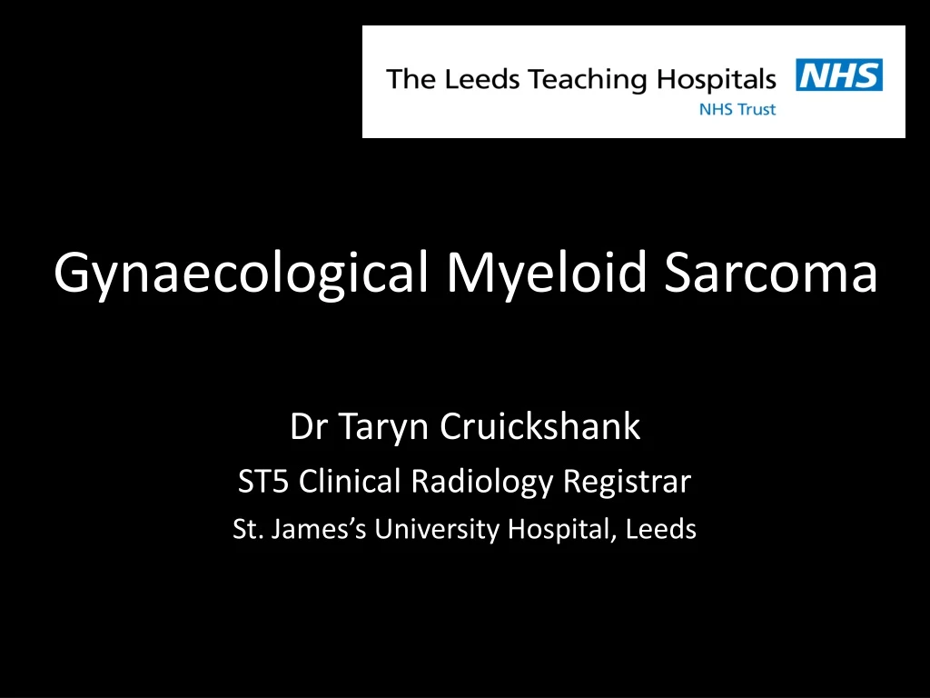
Gynaecological Myeloid Sarcoma Dr Taryn Cruickshank ST5 Clinical Radiology Registrar St. James’s University Hospital, Leeds
Clinical Information A fit and well 71 year old female presented to their GP with a 3 month history of • Lower abdominal pain • Sensation of incomplete bladder emptying • Constitutional symptoms On direct clinical observation, the cervix appeared to be replaced by an indurated mass extending into the anterior and posterior vaginal walls and out to both pelvic sidewalls.
a a a b b b a b Figure 4. a,b. Axial post IV contrast CT imaging demonstrating the gynaecological mass and ureteric stents at presentation (a) and after 6 cycles of chemotherapy (b). * Figure 3. a,b. Avid diffusion restriction of the mass on ADC (a) and B1000 (b) diffusion weighted MRI imaging. Figure 2. a,b. Sagittal (a) and axial (b) T2 MRI images demonstrates a bulky uterine, cervical and vaginal mass with breach of the stromal ring and extension into the parametrium (asterisk). Figure 1. a,b. Ultrasound image (a) demonstrating a large mass positioned posterior to the bladder, indistinguishable from the cervix and uterus. Transvaginal ultrasound guided biopsy (b).
Diagnosis • Blood results on initial presentation demonstrated a high white cell count and an acute renal injury. • The peripheral blood smear demonstrated blasts which led to a new diagnosis of acute myeloid leukaemia (AML). • A transvaginalguided ultrasound core biopsy (figure 1) confirmed a histological diagnosis of myeloid sarcoma.
Key learning points • Myeloid sarcomas can present at any stage of disease, from the first presentation to relapse many years after primary therapy. • On imaging basis, they are mostly misinterpreted as other primary malignancies or lymphoma often due to their overlapping MRI characteristics. • Myeloid sarcomas should be considered in the differential diagnosis of patients presenting with a pelvic mass, with or without a history of myeloproliferative disorder.
Discussion • Myeloid sarcoma (also named granulocytic sarcoma) are extramedullary tumours composed of immature myeloid cells that can present in association with acute leukaemia’s, most commonly acute myeloid leukaemia. 1,2 • The most common locations of myeloid sarcoma include soft tissues, bone, peritoneum, lymph nodes and the gastrointestinal system.2 Cases of extramedullary AML involving the gynaecological organs are exceedingly rare.3 • The patient initially had a a pelvic US and subsequently an MRI and staging CT with IV contrast. The cervix, vagina and the majority of the uterine body were shown to be replaced by tumour with bilateral parametrial involvement and hydronephrosis. Pathologically enlarged nodes were demonstrated in the external iliac groups bilaterally. • The diagnosis is histological, with overlap between the pathological and radiological appearances with lymphoma. • The approach to treating cases of myeloid sarcoma is varied, with management options including a combination of surgery, chemotherapy or radiotherapy.4 In this case, the disease rapidly responded to chemotherapy (figure 4). Abu Saadeh F, Collins V, Al-Saadi M, Gleeson N. An unusual relapse in acute myeloid leukaemia. BMJ Case Rep. 2015;2015:bcr2014207395. Published 2015 Feb 20. Yilmaz AF, Saydam G, Sahin F et al. Granulocytic sarcoma: a systematic review. Am J Blood Res 2013;3:265–70. Garcia MG, Deavers MT, Knoblock RJ et al. Myeloid sarcoma involving the gynaecological tract. Am J Clin Pathol 2006;125:783–90 Weingertner AS, Wilt M, Atallah I et al. Myeloid sarcoma of the uterine cervix as presentation of acute myeloid leukaemia after treatment with low-dose radioiodine for thyroid cancer: a case report and review of the literature. Case Rep Oncol 2009;2:1–6