Download

1 / 26
260 likes | 356 Views
Fluorescence and filters. Phenomenon of fluorescence. Jablonski diagram: Absorption of photon elevates fluorophore to excited singlet state S 1 ’ Nonradiative decay to lowest energy singlet excited state S 1 3. Decay to ground state by emission of a photon. Probes.invitrogen.com.

E N D
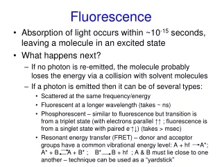
Phenomenon of fluorescence • Jablonski diagram: • Absorption of photon elevates fluorophore to excited singlet state S1’ • Nonradiative decay to lowest energy singlet excited state S1 • 3. Decay to ground state by emission of a photon Probes.invitrogen.com
Everyday examples of fluorescence • Whiteners in fabrics • “Blue hair” "Hey grandma, I dig your blue hair!"
Non-radiative decay to triplet state leads to photbleaching Molecular Expressions website
Ideal fluorophore characteristics • High quantum efficiency • Slow photobleaching • For live cells: excitation wavelengths non-phototoxic to cessl • Little overlap with autofluorescence • Mammalian cells: Flavoprotein, pigment • Plant cells: chlorophyll
A few dyes Probes.invitrogen.com
Fluorescent proteins: GFP Tsien Lab (UCSD)
Fluorescent proteins data table • See http://home.earthlink.net/~pubspectra/
Other useful characteristics of fluorophores – Environmental sensitivity – Fura-2
Ca++-sensitive fluorescent proteins: Cameleons Probes.invitrogen.com
How do we observe fluorescence • Black light • Not enough sensitivity • Filters • Bleed-through • Darkfield fluorescence microscopy
Epifluorescence microscopy Nikon MicroscopyU
Essence of epifluorescence microscope: Dichroic mirror www.microscopyu.com
Dichroic Filters • Fabry-Perot Interferometers: • Constructive interference leads to transmission Molecular Expressions Web Site: Interference filters
Multiple-wavelength interference filters Molecular Expressions Web Site: Interference filters
Excitation filters • Note that Dichromatic mirror has bandpass at short wavelengths Molecular Expressions Web Site: Interference filters
Emission filters • May be either broadband (colored glass) or bandpass • Broadband gives more light • Bandpass filters out more autofluorescence, other fluorophores
Light souces: arc lamps www.microscopyu.com Advantages of mercury: intense illumination at specific wavelengths http://www.olympusmicro.com/primer/anatomy/sources.html
Xenon arcs • More uniform power across visible spectrum http://www.olympusmicro.com/primer/anatomy/sources.html
Dangers of arc lamps • Warm-up period of a new arc lamp • Explosion • Toxic Hg vapors • Solarization of filters • Bleaching of filters • Retinal phototoxicity!!!!
Specimen preparation for fluorescence • Fixation • Formaldehyde • Glutaraldehyde (red autofluorescence) • Coagulation by ice cold methanol plunge • Permeabilization
Chemically specific staining • Histochemistry – stain is a fluorophore • H&E – not specific, not fluorescent • Nissl stain – Nucleic acid spefic, but not fluorescent • DAPI • Chemically labelled toxins • Fluorescent α bungarotoxin • Rhodamine phalloidin
Amanita phalloides Image of Amanita phalloides from Abbé Giacomo Bresadola (1927 - 1960) Iconographia mycologica Death cap mushroom
Indirect immunofluorescence Bradwell A R, Hughes R G, Harden E L (2003)Atlas of HEp-2 patterns, 2nd editionPrinters KNP Group Ltd, Redditch, UKISBN: 0704424371
Our plan • DAPI to detect chromatin • Rhodamine phalloidin to detect f-actin • Monoclonal anti-tubulin (DM1a) to bind to microtubules • Wash • FITC goat-anti-mouse to detect DM1a • Widefield epifluorescence microscopy