Download
1 / 17
230 likes | 717 Views
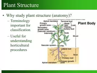
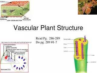
Vascular Plant Structure. Read Pg. 286-289 Do pg. 289 #1-7. http://www.hcs.ohio-state.edu/hcs300/gif/tsstem.gif. Examine the plant you’ve been given. Sketch it and use various resources to label your sketch. Plant Dissection. Parts and Organ Systems of a Flowering Plant.

E N D
Vascular Plant Structure Read Pg. 286-289 Do pg. 289 #1-7 http://www.hcs.ohio-state.edu/hcs300/gif/tsstem.gif
Examine the plant you’ve been given. Sketch it and use various resources to label your sketch. Plant Dissection
Parts and Organ Systems of a Flowering Plant Label using pg. 286
Meristematic Tissue Where cell division occurs Creates all new cells and tissues Differentiation - specialization of cells into certain tissues with certain functions sps.k12.ar.us Tissues http://www.science.smith.edu/departments/Biology/Bio111/xsmith/plants/3tisscut.gif
Waxy Cuticle Protects against damage, water loss and infections Hairs Discourage herbivores Chemicals Irritants Epidermis Outer layer of cells Becomes bark in woody plants Dermal Tissue The Outer Layers
Internal cells Different cell types with different functions e.g. storage, support, photosynthesis E.g. Flesh of fruits and vegetables Ground Tissue The Filler http://www.greentales.com/files/pear.jpg www.greenpatchseeds.com.au http://www.hanung.com/Web/Fruits/Carrot%203119.jpg
Xylem Conducts water and minerals from roots to shoots Thick walled cells Work even when dead Phloem Transports sugar and other solutes from shoots to roots Thin walled cells Living Vascular Tissue Transport
1. body tube 2. rotating nosepiece 3. low power objective lens 4. medium power objective lens 5. high power objective lens 6. stage clips 7. diaphragm 8. light 9. eyepiece (ocular lens) 10. arm 11. stage 12. coarse adjustment knob 13. fine adjustment knob 14. base
Biological Drawing Guidelines • Keep the following guidelines in mind when completing a formal biological drawing of an object you view under the microscope: • 1. Use pencil (preferably soft lead that will not smudge). • 2. Please print. • 3. Please use unlined white 8 1/2” x 11” paper or lab drawing paper. • 4. The drawing should be titled at the bottom with the figure # and appropriate title. Below that you record the magnification. • 5. The drawing should be approximately 1/2 of the page. • 6. Keep the drawing to the left of the center. • 7. Labels are to line up on the right hand side of the page. • -- use a ruler to draw lines from the object to the label • -- lines should never cross over top of each other • –all labels should line up vertically • 8. Do not use plural labels to point to a single object. Keep the labels consistent. • 9. The drawing should be an outline of what you see. Do not include additional structures just because you think you should see them. • 10. Do not shade. All lines should be solid and complete. You may stipple to show different regions. • 11.When using the scientific name of an organism remember that the genus or first part of a scientific name is always capitalized. The species or second part of a scientific name is not. A scientific name is usually italicized as well. • FOR EXAMPLE: Canis familiaris is the genus and species name for a dog.
Activities • Carnation Vascular Tissue Experiment • Viewing slides of stem and leaf with the microscope, label any visible tissues • Hand in: one proper biological diagram of a stem and one of a leaf
http://kentsimmons.uwinnipeg.ca/cm1504/15lab42006/lb4pg4.htm