
Empyema
E N D
Presentation Transcript
Empyema DR. KOMALDEEP JUNIOR RESIDENT PULMONARY MED TBHP
HISTORY • 460 BC hippocratic physicians recommended treating empyema with open drainage. • Napolean’s surgeon dupuytren, whose name is linked to the palmar fascia contractures of liver disease, died of empyema in 1835 after declaring he would “rather die at hands of God than of surgeons” • Sir William Osler who thought “empyema needs a surgeon and 3in. Of cold steel , instead of a fool of a physician” provided a compelling description of his own empyema before succumbing to disease.
THE WORD: EMPYEMA • Em=within • Pyema=accumulation of pus • Empyema is known to be the formation and collection of pus within a naturally present cavity inside the body primarily the pleural cavity • Light’s : “I prefer to reserve the term empyema for those pleural effusions with thick, purulent appearing pleural fluid”.
Other descriptions Weese et al. defined an empyema as pleural fluid with • a sp. Gravity >1.018, • a WBC count >500 cells/mm3 or • a protein level >2.5g/dl. Vianna defined an empyema as pleural fluid on which • the bacterial cultures are positive or • the WBC count is > 15,000/mm3 and • the protein level is >3.0g/dl.
abscess Empyema is different from an abscess because the latter is the formation And collection of pus in a newly formed cavity inside the body. • Parapneumonic effusion: • Any pleural effusion associated with bacterial pneumonia , lung abscess or bronchiectasis is a parapneumonic effusion. • Many complicated parapneumonic effusions are empyemas. • Some patients with empyema have no associated pneumonic process.
Introduction of infection Non-traumatic Traumatic • Direct extension from an adjacent site : lung infection • Aspiration pneumonia • Post-obstructive pneumonia • Bronchiectasis, lung abscess • Instrumentation and rupture of esophagus • Leakage of an esophageal anastomosis after resection • Development of a bronchopleural fistula following pneumonectomy • Pleual aspiration/ tube drainage • Abdominal sepsis: subphrenic abscess , liver abscess • Sepsis in the pharynx, thoracic spine or chest wall may extend into the pleura via tissue planes or mediastinum Non- surgical trauma: • Gun shot wounds, blast injuries and stab wounds.
Pathology (1) Exudative STAGE • Once infected by pathogenic organisms, the connective tissue layers within the pleural memberanes become oedematous and produce an exudation of sterile proteinaceous fluid that starts to fill the pleural cavity. • The deepest layers of the pleural membranes are relatively impervious so that infection tends to be contained within the pleural cavity itself and spread beyond it is unusual. • At this stage, the pleural fluid is thin with a relatively low white cell count and the visceral pleura and underlying lung remain mobile. • Fluid at this stage is having a low WBC count, low LDH level and a normal glucose level and ph.
(2) Fibrinopurulent STAGE • If the infection proceeds unchecked by antimicrobial agents, the inflammatory process continues so that newly formed layers of fibrin become laid down on the epithelial surface within the pleural cavity, particularly on the pleural cavity. • The empyma fluid now becomes more thicker and more turbid, containing, a higher white cell count. • Such empyemas may become loculated into smaller collections by the development of fibrinous bands which prevent the extension of empyema but making the work of percutaneous aspiration difficult or impossible. • With the deposition of fibrin on both pleural surfaces, lung movements in this stage may become increasingly restricted. • The pleural fluid ph and glucose levels becomes progressively lower and LDH level becomes progressively higher.
(3) ORGANIZATIONAL STAGE • Depending upon the nature of the infecting organism and whether or not antibiotics and drainage procedures have been employed, these thickened fibrinous layers organize as collagen and become vascularized by an ingrowth of capillaries. • This stage may begin within two weeks but usually takes 4-6 weeks to develop to a point at which the empyema cavity becomes surrounded by a cortex, peel or rind that may be more than 2 cm thick. • This inelastic pleural peel encases the lung and renders it virtually functionless. • By this time the empyema contains frank pus, which may be viscid.
Ultimately, an inadequately treated empyema cavity may become obliterated and its rind may calcify, producing a so-called firothorax, particularly in case of old tuberculous pleural infection. • The inner layers of the thickened empyema cortex continue to show a considerable inflammatory cell infiltrate and the fibrous outermost layers exert an increasingly restrictive effect, both compressing the underlying lung( the so called “trapped lung” effect) and also tending to draw the overlying ribs together, ultimately producing a chest deformity with a dorsal scoliosis that is concave towards the affected side.
Dry “sicca” pleuritis stage, the inflammatory process of the pulmonary parenchyma extends to the visceral pleura, causing a local pleuritic reaction. • This leads to a pleural rub and a characteristic pleuritic chest pain which originates from the sensitive innervations of the adjacent parietal pleura.
Clinical stages • Acute stage : within the first 2 weeks of the onset. • Chronic Stage : after 2 weeks or with the formation of the thick peel and loculations.
Causes for chronicity • Inadequate Tube Drainage. • Chronic pulmonary Disease( T.B. or Fungal Infection) • Immunosupressed patients. • Presence of Foreign body within the pleural space.
bacteriologic features Influence of pre-disposing factors: • CAP: pneumococcal • HAP: MRSA • Aspiration or lung abscess: anaerobes • Infection from below the diaphragm: gram negative enteric bacilli • External trauma/haemothorax: staph. Aureus • Tuberculous empyema – same mechanism as TB pl. effusion with spillage of large amount of mycobacterium into pleural space purulent effusion that requires surgical intervention and can result in pleural fibrosis and restrictive lung disease
BACTERIOLOGIC FEATURES CONT.. Influence of age: • Anaerobes in elderly • S. pneumoniae in young ambulatory patients • Children: h. influenzae Uncommon microbial causes: fungal(cryptococcus neoformans, blastomyces,coccidioides,histoplasmosis) , actinomyces, nocardia, clostridia, echinococcus spp. , protozoa( trichomonas, entamoeba)
Clinical presentation Aerobic bacterial infections: acute febrile illness Anaerobic bacterial infections: • Subacute illness. • Median symptom duration 10 days • Predisposing factors present : h/o alcoholism, an episode of unconsciousness , poor oral hygeine
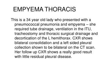
diagnosis • History • Clinical features • Chest radiograph • Ultrasound chest • CT chest • Thoracentesis : empyema fluid : appearance, mirobiology, biochemistry
Chest x-ray • Decubitus view: suspect side down – fluid b/w chest wall and inferior part of lung Suspect side up : parenchymal infiltrate. • In early stages: identical to those of uncomplicated pl. eff. • As the time passes by, fibrosis develops around the empyema cavity so that fluid is contained in one location irrespective of patient’s position. • “D shaped shadow” may be visible along with obliteration of CP angle. • Parenchymal lesion may be visible : consolidation, lung abscess • Air fluid level: pneumothorax : spontaneous or iatrogenic broncho-pleural fistula presence of gas forming organisms such as clostridia
ultrasound • May show septa when there is loculation • Also helpful in targeting an empyema for needle or tube drainage. • Portable • Helpful in distinguishing b/w Loculated pyopneumothorax And Peripheral lung abscess
Computed tomography • Able to detect underlying abnormalities such as oesophageal perforation , bronchial carcinoma and associated lymphadenopathy . • It also aids in the differentiation between empyema and lung abscess. • Empyemas are usually lenticular in shape, compress the lung, and create obtuse angles as they follow the contour of the chest wall. • There is usually an indistinct border between lung parenchyma and a lung abscess, which forms an acute angle where contact with the chest wall is made. • The ‘split pleura’ sign, where both parietal and visceral pleura enhances showing their separation, can be present in an empyema. • Computed tomography (CT) is not as accurate as ultrasound in detecting septations and requires transferring the patient.
Empyema fluid : • GROSS EXAMN : COLOUR, TURBIDITY AND ODOR • Can be distinguished from pleural fluid that is turbid due to chyle i.e. chylothorax by use of centrifugation in which case a whitish layer of chylomicrons is found on surface of pleural fluid. • appearance: E. histolytica: anchovy sauce actinomyces: sulfur granules • Smell: putrid (FECULENT) smell in anaerobic infection.
Microbiology : ZN Gram’s culture: aerobic as well as anaerobic, mycobacterial and fungal • PCR • Cytology : total and differential WBC counts • Ph : should not be done as it will plug up the blood gas machines.
Biochemistry of empyema fluid • Low p H • Low glucose • Raised LDH • Decreased pH and glucose occur as a result of leucocyte and bacterial anaerobic metabolism of glucose and process that produce lactic acid. • Exception The one situation in which pleural fluid ph is not reduced is when the offending organism is of the proteus sp.. These organisms produce ammonia by their urea splitting ability which leads to and elevated pleural fluid ph.
complications • Rupture into the lung: Dissection into lung parenchyma BronchoPleural fistula and pyopneumothorax • Spread to the subcutaneous tissue: Dissection through chest wall EmpyemaNecessitans • Dissection into abdominal cavity. • Septicaemia & septic shock.
Management Principles of management: • Control of the Infection process. • Drainage of pus form the pleura. • Obliteration of the space & complete Re-expansion of the Lung. MANAGEMENT OPTIONS: • General • Medical • Surgery
general • Supportive • Bed rest • Analgesia • Oxygen • Fluids • Identify the cause • Malnutrition • TB • HIV
antibiotic selection • If the fluid’s gram stain and culture reports are available, it should guide the choice of AB. • The initial antibiotic selection : some do not penetrate pleura. • Metronidazole> penicillin>clindamycin>vancomycin>ceftriaxone>gentamicin • Quinolones and clarithromycin also penetrate well 1.Severe CAP : fluoroquinolones(levofloxacin, moxifloxacin, gatifloxacin or gemifloxacin) Advanced macrolide(azithromycin or clindamycin) plus b-lactam(cefotaxime, ceftriaxone) 2. If pseudomonas suspected: piperacilin-tazobactam, imipenem, meropenem 3. Anaerobic: clindamycin or metronidazole. 4. MRSA: vancomycin until culture results are available.
Surgical management “those diseases that medicines do not cure are cured by the knife” TECHNIQUE DEPENDS UPON THE STAGE OF EMPYEMA CLOSED: INTERMITTENT (REPEATED ASPIRATION OR THORACOCENTESIS) CONTINUOUS (INERCOASTAL DRAINAGE ) OPEN: RIB RESECTION ELOESSER FLAP
CLOSED • These methods are more likely to appertain in the exudative stage but may be continued into the fibrino-purulent stage in some cases. • THORACOCENTESIS : frequency with which thoracentesis is repeated depends upon the rate at which pus reaccumulates, which in turn is judged by clinical and radiographic appearance. • Such treatment along with antibiotics is appropriate for many individuals with pleural empyema and these patients may have a shorter and less complicated stay than those by tube drainage.
Closed tube drainage • Tube is placed under local anaesthesia into most dependent part of empyema determined by USG/CT guidance and is connected to an underwater-seal drainage system. • The relatively large (28 to 36F ) tubes have been recommended because of the belief that smaller tubes would become obstructed with the thick fluid. • The advantage of the smaller tube is that it is easier to insert and is less painful to the patient. • Patency is maintained with irrigation and fibrinolytic therapy • If the patient has not demonstrated significant improvement within 24hrs of initiating tube thoracostomy, either the pleural drainage is unsatisfactory(tube placed in wrong position, loculations) or the patient is receiving the wrong antibiotic. • Advantages: successful when infected material is too viscid to remove by manual aspiration. • Disadvantages: greater discomfort and immobility for the patient introduction of new infection at drainage site tube blocked by fibrin clot
Indications for removal of tube: • Volume of the pleural drainage is less than 50ml for 24 hrs and until the draining fluid becomes clear yellow. • The amount of sediment (representing WBCs and debris) in the collection system should not be more than 5ml. • Tube ceases to work Because it serves no useful purpose rather it as a conduit for pleural super-infection.
Closed drainage Successful: • Empyema is small • Is started In acute exudative or early fibrinopurulent stagees of infection In this case, the wall of the empyema cavity gradually becomes absorbed allowing re-expansion of the underlying lung and obliteration of space. It may fail: • If the pus is too thick to drain by thoracentesis or tube • BPF has developed • Pockets of pus become loculated and inaccessible. If drainage is inadequate, ultrasonography or CT should be performed to delineate which factor is responsible. Then, more invasive surgical procedures are required.
Intrapleural streptokinase • The pleural fluid loculations are produced by fibrin membranes that prevent the spread of the infected pleural fluid throughout the body, but which make drainage of the pleural space difficult. • Intrapleural fibrinolytics will destroy the fibrin membranes and facilitate drainage of the pleural fluid. • Indications • Acute or fibrino purulent stage • Presence of loculations. • Incomplete drainage after tube insertion • Contraindications: • Chronic stage • Post-operative empyema • Empyema with BPF.
fibrinolytics • Streptokinase: 15 000U/kg in 20-50ml saline once daily for 3 days (vial 750 000U R1400, 1 million units R2700) • Urokinase: 40 000u in 40ml saline (> 1 year) or 10 000 in 10 ml BD for 3 days(< 1 year) • tPA 0.1mg/kg in 10-30ml saline dwell time 1 hour (50mg vial R3100)
vats • Advantages: • the loculi can be disrupted • pleural space can be completely drained • Chest tube can be optimally placed. • In addition, if lung is trapped(not expanded), the VATS incision can be enlarged so that decortication can be completed with a full thoracotomy.
Pulmonary decortication • In a non-medical aspect, decortication is the removal of the bark, husk, or outer layer, or peel of an object. • It is a surgical procedure that involves the removal of a dysfunctional layer covering the lungs and evacuation of all pus and debris from the pleural space. • The primary aim of performing lung decortication is to be able to promote lung expansion and chest wall compliance. • If after 6months, pleura remains thickened and the patient’s PFTs is significantly reduced to limit activities, decortication should be considered
Open drainage Rib resection: resecting segments of one to three ribs overlying the lower part of empyma cavity and inserting large bore tubes into the empyema cavity. Following this, tubes are irrigated daily with a mild antiseptic solution and daily redressed. Successful open drainage results in gradual obliteration of empyema space. Open window thoracostomy (eloesser flap) : involves the removal of sections of two or more ribs in order to fashion a larger stoma, which is kept open by suturing the skin to the parietal pleura/cortex thereby creating a pleurocutaneous flap, stoma closed if the underlying lung re-expands or may occasionally be left permanently open with daily dressings.
Empyema associated with bpf Adequate pleural drainage is crucial: an emergency • Pleural fluid if not drained exteriorly with chest tubes is likely to drained interiorly into the lung • The bacteria then spread throughout the broncho-pulmonary tree and an overwhelming pneumonia can result. • Drainage should be instituted immediately to prevent the possibility of contaminating the entire respiratory system by the infected pleural fluid. How to suspect: a patient with pleural fluid collection: • Raises more sputum than would be expected • With postural variation • On x-ray(upright position): presence of an air fluid level in the pleural space • Need USG and CT chest to differentiate from a peripheral lung abscess.
Empyema distal to an obstructed bronchus • Contraindication for chest tube placement • If chest tubes placed: bronchial obstruction will prevent the expansion of underlying lung. • What should be done: appropriate AB should be administered along with therapy for obstructed bronchus: radiotherapy, endo-bronchial stent or laser therapy. Once obstruction relieved, ICT to be done.
Post traumatic empyema Factors leading to development of empyema: • Retained hemothorax • Pulmonary contusion • Multiple chest tube placement Management: similar to that of other para-pneumonic effusions.
Post pneumonectomyempyema how to suspect : 2nd day to 7yrs (4wks) • A febrile illness with signs of systemic toxicity • Expectoration of large amounts of pleural fluid • An air-fluid level in the pneumonectomy space • Drainage of purulent material from surgical incision • Mediastinal shift towards contralateral side Organism responsible: staph aureus Diagnosis and treatment : all aptients: AB + chest tube placement
Scheme to move forward • Antibiotics • Pleural aspiration • Chest tube drainage • Chest tube drainage with I/P fibrinolytics • Thoracoscopy • Decortication