Download
1 / 17
170 likes | 182 Views
Unit 1: Cell & Molecular Biology. Cell Growth & Cell Cycle. Learning Intentions. Interphase: G1, S and G2 phases Mitosis: the M phase (4 stages) Cytokinesis Mitotic Index Control of the cell cycle. Abnormal cell division: cancer cells. Four Phases of Cell Division. Cell growth and

E N D
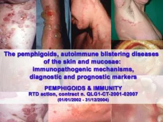
Unit 1: Cell & Molecular Biology Cell Growth & Cell Cycle
Learning Intentions • Interphase: G1, S and G2 phases • Mitosis: the M phase (4 stages) • Cytokinesis • Mitotic Index • Control of the cell cycle. • Abnormal cell division: cancer cells.
Four Phases of Cell Division Cell growth and synthesis of organelles Cell growth and synthesis of organelles Phase G1, S and G2 are collectively known as interphase M phase = Mitosis (cell division) DNA replication
Cell Division And The Cell Cycle Cytokinesis is controlled by actin fibres which split the cytoplasm in two - stage 6
Stages of Mitosis Stage Description Prophase No distinct chromosome. Nuclear envelope intact Prometaphase Chromosome become visible. Nucleus breaks down Metaphase Chromosomes line up across the centre of cell (metaphase plate) Anaphase Chromosomes divide into chromatids which are pulled to opposite poles by spindle fibres. These are made of microtubules and radiate from the centrosome. Telophase Daughter chromosomes ( chromatids) reach opposite poles and begin to de-condense
Interphase Follows the M phase and involves cell growth and DNA replication. Made up of G1, S and G2
Mitosis – Prophase • The replicated chromosomes each consisting of two closely associated sister chromatids condense • Outside the nucleus the mitotic spindle assembles between the two centrosomes which have replicated and moved apart.
Mitosis – Prometaphase • The nuclear envelope breaks down • Chromosomes attach to the spindle microtubules via structures called kinetochores and move towards the equator of the cell.
Mitosis – Metaphase • The chromosomes are moved to the equator by the spindle fibres • The kinetochores of all chromosomes align on the equator, midway between the poles at a structure known as the metaphase plate
Mitosis - Anaphase • The paired chromatids from each chromosome separate to form two sister chromatids. • Daughter chromosomes are pulled to opposite poles by the simultaneous shortening and lengthening of microtubules
Mitosis - Telophase • The two sets of daughter chromosomes arrive at the poles • A new nuclear envelope reassembles around each set forming two separate daughter nuclei and marking the end of Mitosis.
Nuclear division is controlled by microtubules (tubulin protein) which form the spindle fibres and move chromosomes to opposite poles of the cell during mitosis.
Cytokinesis • Once the cell completes M phase (mitosis) the division of the cytoplasm produces 2 daughter cells this is known as CYTOKINESIS.
In animal cells the cytoplasm is divided into two by a contractile ring of actin and myosin protein which pinches in the cell to create two daughter cells. • In plant cells a new cell membrane and cell wall are built between the two daughter nuclei and cuts the cytoplasm in half
Mitotic Index • Mitosis duration and frequency varies greatly between cell types. • The Mitotic index calculates the percentage of cells undergoing division in a given sample. • Only cells goinf through M phase are counted (not Interphase) • This can be the first indication of cancerous cells and a developing tumour.
Mitotic Index = Number of mitotic cells X 100 Total number of cells