Download
1 / 39
390 likes | 726 Views
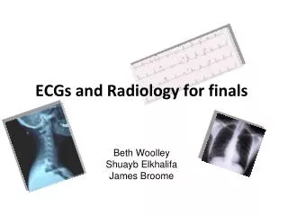
ECGs and Radiology for finals Beth Woolley Shuayb Elkhalifa James Broome. Please present the following ECG trace to the examiner. ECG 1. Qs for ECG 1. Please present this ECG systematically Where was the infarct? Inferior Which artery was affected? Right coronary artery

E N D
ECGs and Radiology for finalsBeth WoolleyShuayb ElkhalifaJames Broome
Qs for ECG 1 • Please present this ECG systematically • Where was the infarct? • Inferior • Which artery was affected? • Right coronary artery • What would the management be in the acute setting? • ABCDE, with the particular emphasis to: • high-flow O2, Venous access, morphine, anti-emetic, GTN, aspirin + clopidogrel. Bloods. CXR. • Primary angioplasty or thrombolysis if no contra-indication 5. What is the management in the longer term? • Modifable RFs. • aspirin (if not contra’d) and a statin, consider ACE-I and B-blocker. • assess need for nitrates and diuretics if in heart failure; cardiac rehabilitation inc. personalised exercise plan.
Qs for ECG 2 • Please present this ECG systematically • RBBB and Q waves (v1-v3) • What are the causes of RBBB? • Can be normal, IHD, cardiomyopathy, and PE, HF • What is the management of RBBB? • Asymptomatic so does not require treatment in its own right • What is a pathological Q wave? Causes? • Likely to be pathological if the Q wave is >2 small squares deep, >25% of the height of the R wave, or >1 small square wide. • Causes of path Q wave: MI, left ventricular hypertrophy and bundle branch block • Where was the old infarct? Which artery? • Antero-septal; left anterior descending artery
Qs for ECG 3 • What is the diagnosis? • What are the common causes of AF? Heart failure, hypertension, cardiac ischaemia, MI, mitral valve disease, pneumonia, hyperthyroidism, alcohol • What are the different components of treatment? • Treat any reversible cause. • Control ventricular rate, e.g. digoxin, B-blocker etc. • Consider cardioversion to sinus rhythm if heart is structurally normal. • Prevent emboli: aspirin or warfarin
Mr X is a 78 year old lady who presented to the accident and emergency department after a fall on ice. She reports falling to her right. On examination she is tachycardic, clearly in pain and her right lower limb is externally rotated. Please discuss your acute management plan.
Whats the diagnosis? • Whats the acute Mx? • ABCDE. • A – Patent • B- Clear? Sats and RR? • C-BP? HR? UO? Shocked. Tx accordingly • D – GCS? BM? PEARL? • Head Injury? Tx accordingly • E - Abdo SNT? • Other fractures? • provide analgesia. • Assess the fracture:
Assessing the #NOF Shortened and externally rotated (if displaced) Pain on ext or int rotation Pain on loading Neuro status? Vascular compromise? Other Inv: FBC, UE, Glu, G&S, Exclude medical causes for fall: ECG, ?CXR. Orthopaedic input
Mrs Jones is a 67 year old lady who tripped on a loose rug. She fell forward and attempted to stop her fall with both hands. • On entering A&E her Left arm in a sling. • You order a plain film of the left arm, and the orthopaedic registrar asks you to describe the film over the phone.
Describe the fracture: This is a AP and lateral film of Mrs Jones left arm. The most striking abnormality is a transverse fracture of the distal radius The distal segment is dorsally angulated and laterally displaced. It is a simple fracture and closed.
Describing fractures Opening AP/PA film Patients Name Area (i.e. L arm) Date of study. Site Bone affected Proximal / Distal Direction of # Line Transverse - Longitudinal | Oblique / Spiral ) • Relationship of distal fragment: • Displaced (lateral / medial) • Angulated (volar / dorsal, A/P) • Shortened • Rotated • No. of fragments • 2 = simple • >2 = comminuted • Soft Tissues • Open / closed • Best evaluated clinically
Mr Harris is a 72 year old gentleman who sustained a fall from a 5 foot ladder. He reports an irregular heart rhythm for which he takes warfarin. • He has a reduced GCS (14) and has been vomiting. His wife reports he was initially fine. • What imaging modalities would you request as part of you management plan • Describe the CT findings.
This is a non-contrast enhanced head CT of Mr Harris. The most striking abnormality is an hyperdense cresenteric mass in the overlying the Right parietal lobe. There also appears to be mass effect, represented by the midline shift. These findings are consistent with a subdural haematoma. How would you manage this acutely? ABCDE
85 yrs, male P/C cough and left sided chest pain for 6weeks .. Qs: 1- present the CXR? 2- Diagnosis? 3- Plans of management?
A single, 3cm relatively thin-walled cavity is noted in the left midlung. This finding is most typical of squamous cell carcinoma (SCC). One-third of SCC masses show cavitation
Management • Staging CT scans. • Surgery/Chemotherapy/ Radiotherapy. • Poor prognosis.
Example Of … Metastatic Lung Cancer: multiple nodules seen
54 yrs, male, smoker 30 CPD, admitted with worsening SOB and cough for 2 days. 1- Describe CXR. 2- Diagnosis and management.
COPD: flattening of the diaphragm, and increase in the size of the retrosternal air space. In addition the upper lobes will become hyperlucent due to destruction of the lung tissue. Management: pred + nebs+/- antibiotics
60 yrs, female, admitted with worsening SOB and Cough for last one week. ( using 3 pillows at night) Qs: 1- Describe CXR? 2- Diagnosis? 3- Management?
CHF:a great deal of accentuated interstitial markings, Curly lines, and an enlarged heart. Normally indistinct upper lobe vessels are prominent but are also masked by interstitial edema. • Management: Diuretics, monitor U&Es
42 yrs, male, was admitted with Cough, fever and night sweats. Qs: 1- Describe CXR ? 2- Diagnosis? 3- Management?
Upper lobe consolidation, consistent with TB. Management: Standard 6 months TB regimen: 6 months:isoniazid and rifampicin And for the first 2 months: pyrazinamide & ethambutol
35 yrs, male, admitted with worsening SOB, cough and Left sided chest pain for one week: 1- Describe CXR? 2- Diagnosis? 2- Management plans?
Pleural effusion: Note loss of left hemidiaphragm. Fluid drained via thoracentesis. Management: • Pleural tap/ sample. (3 samples) • Light’s criteria.
Lights criteria Exudative pleural effusions meet at least one of the following criteria, whereas transudative pleural effusions meet none: 1. pleural fluid protein/serum protein >0.5 2. pleural fluid LDH/serum LDH >0.6 3. pleural fluid LDH more than two-thirds normal upper limit for serum