Download
1 / 69
700 likes | 1.04k Views
Auditory and Vestibular System. http://www.hearingcarecenter.com. Much of the text material is from, “Principles of Anatomy and Physiology, 14th edition” by Gerald J. Tortora and Bryan Derrickson (2014). I don’t claim authorship. Other sources are noted when they are used.

E N D
Auditory and Vestibular System http://www.hearingcarecenter.com
Much of the text material is from, “Principles of Anatomy and Physiology, 14th edition” by Gerald J. Tortora and Bryan Derrickson (2014). I don’t claim authorship. Other sources are noted when they are used. Mappings of the lecture slides to the 12th and 13th editions are provided in the supplements.
Outline • Introduction • Basic structure • Nature of sound waves • Physiology of hearing • Physiology of equilibrium
Overview • The ear contains sensory receptors sensitive to sound vibrations of extremely small amplitudes (as little as 0.2 nm). • These receptors can respond 1,000 times faster than photoreceptors sensitive to light. • The ear has vestibular receptors to maintain the body’s equilibrium and balance. Vestibular = relating to the perception of body position and movement. (from www.merriam-webster.com) Equilibrium = a state of bodily balance, maintained primarily by special receptors in the inner ear. (from www.thefreedictionary.com) Chapter 17, page 595
Sounds Surround Us http://www.potential2success.com www.panoramio.com http://www.ascienceportal.com http://whatbird.wbu.com http://avrackradio.webs.com
Vestibular Sensations Can Challenge Us http://sciencefair.math.iit.edu http://phinn.funformentals.com
Major Components of the Ear • The ear has three major components: • The external or outer ear collects sound waves and channels them inward. • The middle ear conveys sound waves to a structure known as the oval window. • The internal ear has sensory receptors for hearing, equilibrium, and balance. Figure 17.18 Chapter 17, page 595
Cross-Section of the Ear Auricle http://www.health.state.ny.us
External Ear • The external ear consists of the auricle, external auditory canal, and tympanic membrane or eardrum. • The auricle is a flap of elastic cartilage, shaped like the flared end of a trumpet and covered by skin. • The external auditory canal is a curved tube about 2.5 cm in length that lies in the temporal bone, and leads to the tympanic membrane. • The tympanic membrane (ear drum) is a thin, semi-transparent partition located between the external auditory canal and middle ear. Figure 17.18 Chapter 17, page 595
Tympanic Membrane Damage • A tear in the tympanic membrane is known as a perforated eardrum. • The damage can result from a foreign object, trauma, or middle ear infection. • The old adage applies, “never put anything in your ear smaller than your elbow.” • Fortunately, a perforated eardrum will typically heal in about one month. Chapter 17, page 595
An otoscope—a viewing instrument for illumination and magnification— is used to observe the external auditory canal and tympanic membrane. http://graphics8.nytimes.com Chapter 17, page 595 Otoscope
Cerumen • Ceruminous glands are specialized sweat glands that secrete ceru-men, or earwax. • The combination of cerumen and hairs in the external auditory canal helps prevent dust and foreign objects from entering the ear. • Cerumen also helps prevent damage to the skin of the external aud-itory canal by water and insects. Chapter 17, page 596
Cerumen (continued) • While cerumen usually dries-up and falls out, some people produce large amounts of earwax that become impacted and muffle sounds. • Treatment by medical personnel involves irrigation of the external auditory canal, or removal of the build-up with a blunt instrument. http://ukweli.files.wordpress.com Chapter 17, page 596
Middle Ear • The middle ear is a small, air-filled cavity lined by epithelium and located in the temporal bone. • It is separated from the external ear by the tympanic membrane, and from the inner ear by the oval window and round window. Figure 17.19 Chapter 17, page 596
Middle Ear (continued) http://www.phon.ox.ac.uk
Auditory Ossicles • The three smallest bones in the body, called the auditory ossicles, are the major parts of the middle ear, and are attached to it by ligaments. • The bones, which have Latin names for their shapes, are the malleus, incus, and stapes. • The three bones are also known as the hammer, anvil, and stirrup. • The stapes, or stirrup, and its footplate fit into the oval window of the inner ear. Figure 17.19 Chapter 17, page 596
Tensor Tympani Muscle • Two very small skeletal muscles are attached to the auditory ossicles. • The tensor tympani muscle, supplied by the trigeminal (V) cranial nerve, limits the movement of the auditory ossicles. • Muscle contraction increases the tension on the tympanic membrane to reduce damage from loud sounds. Figure 17.19 Chapter 17, page 596
Stapedius Muscle • The stapedius muscle, supplied by the facial (VII) cranial nerve, dam-pens large vibrations of the stapes to protect the oval window. • The damping by the stapedius muscle reduces hearing sensitivity in response to loud sounds. Damping = the capacity built into a device to prevent excessive correction and the resulting instability or oscillatory conditions. (from www.answers.com) Figure 17.19 Chapter 17, page 596
Muscle Response • The tensor tympani and stapedius muscles require only a fraction of a second to contract. • The muscles help protect the inner ear from some loud noises, but not from very brief sounds such as gunfire. • Paralysis of the stapedius muscle can result in hyperacusia, or abnor-mally-sensitive hearing. Chapter 17, page 596
Auditory Tube • The middle ear has an opening that leads to the auditory tube, which is also known as the eustachian tube. • The tube, composed of bone and cartilage, leads to the nasopharynx, the superior portion of the throat. • The auditory tube is normally closed at the pharyngeal or throat end. • It opens during swallowing and yawning, enabling air to enter or exit the middle ear until the pressure in the middle ear equals atmospheric pressure. • This process is known as as equilibration. Figure 17.19 Chapter 17, page 597
Auditory Tube (continued) • When the pressures are equalized, the tympanic membrane can vi-brate freely when sound waves strike it. • Intense pain, hearing impairment, ringing sensations, and vertigo can develop if the pressures are not equalized. • The auditory tube can serve as a path for pathogens from the nose and throat to the middle ear that can cause ear infections. Vertigo = aside from being the name of a classic 1958 Alfred Hitchcock film (with Jimmy Stewart and Kim Novak), vertigo is a feeling that you are dizzily turning around or that things are dizzily turning about you. Vertigo is usually due to a problem with the inner ear. Vertigo can also be caused by vision problems. (from www.medterms.com) Chapter 17, page 597
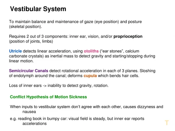
Inner Ear • The internal or inner ear is also known as the labyrinth—it consists of an outer bony structure that encloses an inner membranous labyrinth. • This bony structure, known as the bony labyrinth, has cavities in the temporal bone that are divided into three areas: • Cochlea with receptors for hearing • Vestibule with receptors for static equilibrium • Semicircular canals with receptors for dynamic equilibrium Membranous = relating to or made of a membrane. Figure 17.20 Chapter 17, page 597
Inner Ear (continued) http://rivercitymalone.com
Vestibule • The vestibule is the oval central portion of the bony labyrinth positioned between the semicircular canals and the cochlea. • It contains two sacs, known as the utricle and saccule, to form the mem-branous labyrinth. • Three semicircular canals project from the vestibule at approximate right angles to each other. Figure 17.20 Chapter 17, page 597
Semicircular Canals • The three structures are known as the anterior, posterior, and lateral semicircular canals. • The first two have a vertical orientation, and the third has a horizontal orientation. • The enlargement at one end of each semicircular canal is known as the ampulla. • The membranous labyrinth within the semicircular canals is known as the semicircular ducts, which connect with the utricle. Figure 17.20 Chapter 17, page 597
Vestibular Innervation • The vestibular branch of the vestibulocochlear (VIII) cranial nerve has first-order sensory neurons and motor neurons. • First-order sensory neurons transmit sensory information from the ves-tibular receptors to the brain. • Motor neurons carry feedback signals (action potentials) from the brain to the vestibular receptors to modify their sensitivity to linear and rota-tional accelerations. Innervation = supply an organ or body part with nerves. Chapter 17, page 598
Cochlea • The cochlea is a spiral canal anterior to the vestibule—the canal resembles a snail’s shell, and makes almost three turns around its bony core. • The cochlea has three channels—cochlear duct, scala vestibuli, and scala tympani. Figure 17.21 Chapter 17, page 598
Cochlea (continued) • The vestibular membrane separates the cochlear duct from the scala vestibuli. • The basilar membrane separates the cochlear duct and the scala tym-pani. • The cochlear duct is filled with endolymph, and the scala vestibuli and scala tympani are filled with perilymph. Endolymph = the fluid that fills the membranous labyrinth of the internal ear. Perilymph = the fluid that surrounds the membranous labyrinth of the inner ear. Figure 17.21 Chapter 17, page 598
Cochlea (continued) • The spiral organ, or organ of Corti, rests on the basilar membrane of the inner ear. • It contains a coiled sheet of epithelial cells with about 16,000 hair cells that serve as the receptors for hearing. • The inner hair cells are arranged in a single row, and the outer hair cells are arranged in three rows. Figure 17.21 Chapter 17, page 598
Stereocilia • About 40-to-80 stereocilia at the tip of each hair cell extend into the endo-lymph of the cochlear duct. • The tectorial membrane, a flexible gelatinous (gelatin-like) tissue, covers the hair cells of the spiral organ. Electron micrograph of stereocilia in a frog http://www.unmc.edu Chapter 17, page 598
Cochlear Innervation • The inner and outer hair cells synapse with first-order sensory neurons and motor neurons from the cochlear branch of the vestibulocochlear (VIII) cranial nerve. • As mentioned, the outer hair cells outnumber inner hair cells by a factor of 3-to-1. • The inner hair cells, however, synapse with 90-to-95 percent of the first-order sensory neurons that relay auditory information to the brain. • About 90 percent of the motor neurons, however, synapse with outer hair cells. Figure 17.21 Chapter 17, page 598
Sound Waves • Sound waves are alternating higher- and lower-pressure regions that travel through a medium such as air or water. • They originate from a vibrating object in much the same manner that ripples travel over the smooth surface of a lake when a stone is tossed into it. http://www.zgeek.com Chapter 17, page 598
Sound Frequencies • The frequency of a sound wave is known as its pitch—the higher its frequency, the higher the pitch. • The entire audible range for young adults extends from 20 to 20,000 Hz. • Humans typically have their greatest auditory sensitivity between 50 and 5000 Hz. • Speech sounds are usually in the range between 100 and 3000 Hz. • The audible range diminishes as a person progresses into middle age and older. Chapter 17, page 598
Some Reference Frequencies • Middle C = 261.6 Hz, and a high C sung by a soprano = 1048 Hz. • Tuning of modern musical instruments, A above middle C = 440Hz. Queen of the Night—Mozart’s The Magic Flute http://www.wga.hu Chapter 17, page 598
Sound Intensity • Intensity is the amplitude of a vibration—we perceive it as the loudness of a sound. • Sound intensity is measured in decibels (dB), which are measured on a logarithmic scale. • An increase of one dB represents a ten-fold increase in sound intensity. Chapter 17, page 598
Sound Intensity (continued) • Typical dB levels are: • Gently rustling leaves, 15 dB • Whispered speech, 30 dB • Normal conversation, 60 dB • Vacuum cleaner, 75 dB • Shouting, 80 dB • Nearby loud motorcycle, 90 dB • To a healthy ear, sounds become uncomfortable at 120 dB, and painful above 140 dB. Chapter 17, page 598
Decibel Comparisons http://www.cyberphysics.co.uk
Sequence of Events The auricle directs sound (pressure) waves into the external auditory canal. The sound waves strike the tympanic membrane causing it to move back-and-forth—the eardrum vibrates slower for low-frequency than high-frequency sounds. The vibration is transmitted from the malleus (connected to the central area of the tympanic membrane), to the incus, and then to the stapes. Figure 17.22 Chapter 17, page 601
Sequence of Events (continued) As the stapes moves back and forth, it pushes the membrane of the oval window in-and-out—it vibrates more intensely than the tympanic membrane due to its smaller size. Movement of the oval window creates pressure waves in the perilymph of the scala vestibuli in the cochlea. The pressure waves are transmitted from the scala vestibuli to the scala tympani, and then to the round window. Figure 17.22 Chapter 17, page 601
Sequence of Events (continued) The pressure waves that deform the walls of the scala vestibuli and scala tympani push the vestibular membrane back and forth, which produces pressure waves in the endolymph inside the cochlear duct. The pressure waves in the endolymph cause the basilar membrane to vibrate, moving the hair cells of the spiral organ against the basilar membrane. The movements result in bending of the stereocilia, producing graded, receptor potentials that lead to the generation of action potentials. The action potentials propagate along the cochlear branch of the vest-ibulocochlear (VIII) cranial nerve to the brain, as will be covered in sub-sequent slides. Figure 17.22 Chapter 17, page 601
Frequency Tuning • Sound waves of differing frequencies (pitch) cause different segments of the basilar membrane to vibrate more effectively than other segments. • Each segment of the basilar membrane is tuned for a specific frequency. • The basilar membrane is narrower and stiffer at the base of the cochlea, and therefore high-frequencies induce maximal vibrations in this region. • The basilar membrane is wider and more flexible toward the apex (tip) of the cochlea, and therefore low-frequencies induce maximal vibrations in this region. Chapter 17, page 602
Cochlear Organization Apex Base http://www.physics.uwo.ca
Sensitivity to Sound Intensities • The loudness we perceive is determined in large part by the intensity, or amplitude, of the sound waves. • High-intensity sounds produce larger vibrations in the basilar membrane, resulting in a higher frequency of action potentials in the cochlear branch of the vestibulocochlear nerve. • Louder sounds also stimulate a larger number of hair cells in the cochlea. Chapter 17, page 602
Transduction • The hair cells in the basilar membrane transduce mechanical vibrations into electrical signals. • As the basilar membrane vibrates, the stereocilia at the apex of the hair cell bend back and forth, and slide against one another. • The bending of the stereocilia produces graded, receptor potentials via a tip link protein that connects the tip of each stereocilium with a mechani-cally-gated ion channel. • Depolarization results from the inflow of K+ from the endolymph, produc-ing an inflow of Ca2+ that triggers synaptic vesicles to release glutamate, a neurotransmitter. Transduce = to convert energy from one form to another. Chapter 17, page 602
Transduction (continued) • With Ca2+ inflow, the frequency of the action potentials increases in first-order sensory neurons that synapse with the base of the hair cell. • Bending of the stereocilia in the opposite direction generates hyper-polarizations, which decreases the frequency of the action potentials in the first-order sensory neurons. Chapter 17, page 602
Otoacoustic Emissions • The cochlea can produce sounds n its own although they are usually inaudible. • These otoacoustic emissions are recorded by placing a special micro-phone next to the tympanic membrane. • The emissions are produced by the vibrations of the outer hair cells in response to signals from motor neurons that innervate the cochlea. • The activity enhances the movement of the basilar membrane, which serves to amplify the response of inner hair cells to sound waves. Chapter 17, page 602