Download
1 / 81
910 likes | 1.73k Views
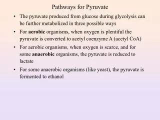
Pyruvate Carboxylase. Reversing the final steps. Inverse regulation of glycolysis and gluconeogenesis. Hexokinase IV in liver Hexokinase I in muscle. HxkIV Km = 10 mM. When blood glucose drops below 5 mM, F6P inhibits it. This way liver does not compete with muscle for glucose.

E N D
Hexokinase IV in liver Hexokinase I in muscle HxkIV Km = 10 mM
When blood glucose drops below 5 mM, F6P inhibits it. This way liver does not compete with muscle for glucose
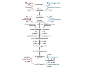
Electron transport and oxidative phosphorylation Glucose is completely oxidized to CO2 through the enzymatic reactions of glycolysis and the citric acid cycle. The redox equation for this process is: C6H12O6 + 6O2 ---> 6CO2 + 6H2O ΔG°’ = -2823 kJ.mol-1 Which can be represented by two half reactions: C6H12O6 + 6H2O ---> 6CO2 + 24H+ + 24e- glucose is oxidized 6O2 + 24H+ + 24e- ---> 12H2O molecular oxygen is reduced In living systems the electron transfer process connecting these two half reactions occurs through a multistep pathway that harnesses the liberated free energy to form ATP.
The 12 electron pairs involved in glucose oxidation are not transferred directly to O2. Rather they are transferred to coenzymes NAD+ and FAD to form NADH and FADH2 10 NADH : 20 e- 2 FADH2 : 4 e- The sites of electron transfer that form NADH and FADH2 in glycolysis and the citric acid cycle are represented in the figure.
The electrons are extracted from the cofactors by reoxidation and then join the electron-transport chain, in this process, protons are expelled from the mitochondrion. The free energy stored in the resulting pH gradient drives the synthesis of ATP from ADP and Pi (inorganic phosphate) through oxidative phosphorylation. Reoxidation of NADH ~ 3 ATP Reoxidation of FADH2 ~ 2 ATP A total of 38 ATP are produced per each molecule of glucose completely oxidized to CO2 and H2O (including the 2 ATP made in glycolysis and the 2 ATP made in the citric acid cycle)
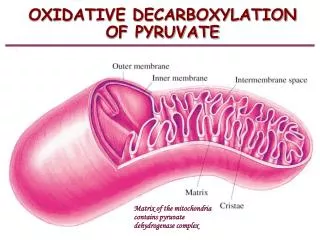
Mitochondria is the site of eukaryotic oxidative metabolism 0.5 m in diameter and 1 m long (about the size of a bacterium) The outer membrane contains porin, a protein that forms pores and allows free difussion of up to 10 kD molecules The inner membrane is a lot more dense and is permeable only to O2, CO2 and H2O. Contains numerous transport proteins that control metabolite passage.
Mitochondrion is not a regular shaped organelle it is a dynamic organelle that is reticulated throughout the cell
Electrons enter the electron transport chain onto Q Glycerol phosphate shuttle NADH:Q Oxidoreductase Succinate dehydrogenase Fatty acid metabolism
The glycerophosphate shuttle mainly occurs in rapidly metabolizing tissues.
NADH from glycolysis are generated in cytoplasm Problem: No way to transport NADH into the mitochondrion to be reoxidized! Solution: Use the malate-aspartate shuttle
Complex I: NADH:CoQ oxidoreductase NADH + H+ + CoQ(ox) + 4H+(in) ---> NAD+ + CoQH2(red) + 4H+(out) ∆E = 0.360 V ∆G = -69.5 kJ/mol -0.030 +0.045 -0.25 -0.30 -0.32
-0.32 -0.30 Ubiquinone +0.045
Complex II: succinate dehydrogenase Succinnate:CoQ oxidoreductase FADH2 + CoQ(ox) ---> FAD + CoQH2(red) ∆E = 0.085 V ∆G = -16.4 kJ/mol
-0.031 -0.040 -0.030 -0.245 -0.060 -0.080 +0.045
-0.031 Succinate
Complex III CoQH2(red) + 2cyt c(ox) + 2H+(in) ---> CoQ(ox) + 2cyt c(red) + 4H+(out) ∆E = 0.190 V ∆G = -36.7 kJ/mol
+0.235 +0.215 +0.280 +0.045 -0.030 +0.030
CoQH2 + cyt c1(ox) ---> CoQ•- + cyt c1(red) + 2H+(out) Cycle I CoQH2 + CoQ•- + cyt c1(ox) + 2H+(in) ---> CoQ + CoQH2 + cyt c1(red) + 2H+(out) Cycle II CoQH2 + 2cyt c1(ox) + 2H+(in) ---> CoQ + 2cyt c1(red) + 4H+(out) Net Reaction 4 protons pumped instead of 2
Complex IV 4 cytochrome c2+ + O2 + 8H+(in) => 4 cytochrome c 3+ + 2H2O + 4H+(out) ∆E = 0.580 V ∆G = -112 kJ/mol
+0.235 +0.245 +0.815 +0.385 +0.340
If 2 electrons enter at complex I 4 + 4 + 2 = 10 protons pumped out If 2 electrons enter at complex II or Glycerol dehydrogenase or fatty acid metabolism 4 + 2 = 6 protons pumped out