Download
1 / 65
660 likes | 816 Views
Ling 411 – 07. Brain Damage and Locations of Linguistic Functions. Why so much variation in symptoms?. Difference in areas of brain damage Difference in kinds of brain damage Strokes vs trauma vs infection vs tumors Different kinds of stroke Anatomical variation among people

E N D
Ling 411 – 07 Brain Damage and Locations of Linguistic Functions
Why so much variation in symptoms? • Difference in areas of brain damage • Difference in kinds of brain damage • Strokes vs trauma vs infection vs tumors • Different kinds of stroke • Anatomical variation among people • Differing cortical structures • Differences in vascular anatomy • Difference in location of cortical functions
Why so much variation in symptoms? • Difference in areas of brain damage • Difference in kinds of brain damage • Strokes vs trauma vs infection vs tumors • Different kinds of stroke • Anatomical variation among people • Differing cortical structures • Differences in vascular anatomy • Difference in location of cortical functions
Different types of brain damage • Strokes, wounds, tumors, infections, degenerative disease • Each of these occurs in varying locations • Each of these has varying extent of damage
Different Kinds of Stroke Damage • Ischemic: blockage of artery • Two sources of blockage: • Thrombosis (about 2/3 of all ischemic strokes) (B&A 64) • Embolism: caused by a blood clot, air bubble, or detached clot • Result: infarction – death of brain tissue that is no longer receiving blood supply • Variation in location of blockage • Hence, variation in area of infarction • Hemorrhagic: bleeding into cerebral tissues • Variation in location and extent of hemorrhage
Stroke mechanisms http://www.youtube.com/watch?v=M_fo6ytlmD0&feature=related
Why so much variation in symptoms? • Difference in areas of brain damage • Difference in kinds of brain damage • Strokes vs trauma vs infection vs tumors • Different kinds of stroke • Anatomical variation among people • Differing cortical structures • Differences in vascular anatomy • Difference in location of cortical functions
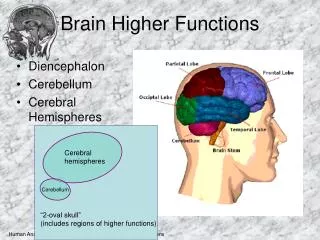
Cerebral Arteries • Anterior Cerebral Artery • Feeds frontal pole and most of the medial surface • Middle Cerebral Artery • Feeds most of cortex, • Perisylvian area • Other areas • Posterior Cerebral Artery • Feeds bottom of temporal lobe and medial surface of occipital and parietal lobes
View of actual brain with arteries http://www.youtube.com/watch?v=Qn4NArz385U&feature=related
Middle Cerebral Artery (Right Hemisphere) www.strokecenter.org/education/ais_vessels/ais049b.html The middle cerebral artery is the largest branch of the internal carotid. The artery supplies a portion of the frontal lobe and the lateral surface of the temporal and parietal lobes, including the primary motor and sensory areas of the face, throat, hand and arm and in the dominant hemisphere, the areas for speech. The middle cerebral artery is the artery most often occluded in stroke. From Washington University Medical School
Territory Anterior cerebral artery occlustion Posterior cerebral artery occlusion Middle cerebral artery occlusion Aphasic syndrome Extrasylvian motor aphasia Occipital alexia Various major types of aphasia (next slide) Aphasic syndromes and Cerebrovascular areas
Total artery occlusion Orbitofrontal branch Rolandic branch Anterior parietal branch Posterior parietal branch Angular branch Posterior temporal branch Anterior temporal branch Global aphasia Broca aphasia Broca aphasia, cortical dysarthria Conduction aphasia Wernicke aphasia, extrasylvian sensory aphasia Anomia, extrasylvian sensory aphasia Wernicke aphasia Anomia Aphasias with middle cerebral artery occlusion
Why so much variation in symptoms? • Difference in areas of brain damage • Difference in kinds of brain damage • Strokes vs trauma vs infection vs tumors • Different kinds of stroke • Anatomical variation among people • Differing cortical structures • Differences in vascular anatomy • Difference in location of cortical functions
Neuroanatomical correlates of the aphasiasIdentifying linguistic functionsLocating linguistic functions
Evaluating evidence from aphasia • It would be easy if naïve localization were true • If a patient has lost an ability, then the area of damage is the area responsible for that ability • But naïve localization is false • “… language, along with other complex cognitive processes, depends on the concerted operation of multicomponent, large-scale neural systems. The anatomical components are often widely dispersed and each acts as a partial contributor to a complicated process…” Antonio Damasio 1998:25
Simple Functions / Complex Functions Complex function Simple function Suppose this area gets knocked out. Then the whole complex function is impaired
Benson and Ardila on conduction aphasia “… a single type of aphasia may have distinctly different loci of pathology. Both conduction aphasia and transcortical motor aphasia are examples of this inconsistency.” (117) (See also p. 135)
Hannah Damasio on conduction aphasia “Conduction aphasia is associated with left perisylvian lesions involving the primary auditory cortex…, a portion of the surrounding association cortex…, and to a variable degree the insula and its subcortical white matter as well as the supramarginal gyrus (area 40). Not all of these regions need to be damaged in order to produce this type of aphasia. In some cases without involvement of auditory and insular regions, the compromise of area 40 is extensive…. In others, the supramarginal gyrus may be completely spared and the damage limited to insula and auditory cortices … or even to the insula alone….” (1998: 47)
CT template – Conduction Aphasia (patient II) Left auditory cortex and insula
MR template – Wernicke Aphasia (patient I) Poster-ior portion of super-ior and middle temp-oral gyri
MR template – Wernicke Aphasia (patient II) Super-ior temp-oral gyrus, AG, SMG
CT template – Broca Aphasia (patient I) Superior sector of Broca’s area and the pre-motor region immedi-ately above it
MR template – Broca Aphasia (patient II) Most of Broca’s area, motor and pre-motor regions, white matter, insula
MR template – Transcortical Motor Aphasia Motor and pre-motor cortices just above Broca’s area
Two different patients with anomia Inability to retrieve words for unique entities (Left temporal lobectomy) Deficit in retrieval of animal names (Damage from stroke)
Two more patients with anomia Deficit in retrieval of words for man-made manipulable objects (Damage from stroke) Severe deficit in retrieval of words for concrete entities (Herpes simplex encephalitis)
More on these four anomic patients • “…anomic aphasia requires damage to left temporal cortices located outside the traditional aphasia-producing territories” (Hannah Damasio 1998:50) • All of these four subjects demonstrated normal concept retrieval for the concrete entities they could not name (Hannah Damasio 1998:51) (But she does not elaborate – maybe it’s not really “normal” – possibly RH conceptual knowledge?)
Don’t forget this – (repeating) • Some information is bilaterally represented • Highly entrenched items • Initial consonants of high-frequency words (?) • Some people have more bilateral representation than others • Women and left-handers tend to have more bilateral representation than men and righties • Semantic information is in both LH and RH • But different aspects of semantic information • Metaphor, irony, sarcasm, pragmatic features, inferencing, subserved by RH
Conceptual category dissociation I(from Rapp & Caramazza 1995) • J.B.R. and S.B.Y. (905b-906a) • Herpes simplex encephalitis • Both temporal lobes affected • Could not define animate objects • ostrich, snail, wasp, duck, holly • Much better at defining inanimate objects • tent, briefcase, compass, wheelbarrow, submarine, umbrella • How to explain?
Conceptual category dissociation II • J.J. and P.S. (Hillis & Caramazza 1991) (906-7) • J.J. – left temporal, basal ganglia (CVA) • Selective preservation of animal concepts • P.S. – mostly left temporal (injury) • Selective impairment of animate category P.S J.J.
MR template – Transcortical Sensory Aphasia AG and post-erior SMG
Transcortical sensory aphasia(A. Damasio 1998:36) • Fluent and paraphasic speech • Global paraphasias • Severe impairment in oral comprehension • Repetition intact (unlike Wernicke’s aphasics) • N.b.: Refers to H. Damasio, Chapter 3, for localization of damage
Brain damage and nominal concepts • Access to nominal concepts is impaired in extra-sylvian sensory aphasia • Type I – Damage to temporal-parietal-occipital junction area • I.e., lower angular gyrus and upper area 37 • Poor comprehension • Naming strongly impaired • Semantic paraphasia • Type II – Damage to upper angular gyrus • Variable ability to comprehend speech • Naming strongly impaired • Few semantic paraphasias • Many circumlocutions Benson & Ardila 1996
Summary: Correlations of symptomswith areas of lesion Aphasic Syndrome Area of Damage Cf. H. Damasio 1998: 43-44
Correlation of aphasia types to localization of damage “More than 100 years of study of anatomoclinical correlations, with autopsy material as well as CT and MR scans, has proven that in spite of the inevitable individual variability, the correlation between aphasia types and locus of cerebral damage is surprisingly consistent.” Hannah Damasio 1998: 64
Correlation of linguistic functions to localization of aphasic damage “…the correlations per se provide only limited information about the neurobiological mechanisms of language, in health and in disease.” Hannah Damasio 1998: 64-6
Reasoning from brain damage to localization • If area A is damaged and patient has deficit D of some function F • Does this mean that function F is subserved by area A? • Not really.. • It means that A (or some portion of A) is needed for some component of F
Brain damage and localization of functionHypothetical example A function Damage
Alexia and Agraphia • Alexia with agraphia • Reading and writing both impaired • A rare disorder • Patients with both impairments usually also have Wernicke’s aphasia or transcortical sensory aphasia • Alexia without agraphia, a.k.a. pure alexia • Reading impaired, writing okay • Can write spontaneously or to dictation • Some can copy writing but with difficulty
Misprint in Antonio Damasio Reading • Antonio Damasio, Signs of Aphasia • P. 38: “As the designation implies, patients presenting alexia with agraphia become unable to read while they continue to be able to write…” • Should be “…alexia without agraphia…”
More on patient J.G. • Damage: Left posterior temporal-parietal • Meaning spared, phonological recognition okay, but couldn’t spell the word correctly • digit: • D-I-D-G-E-T • “A number” • thief: • T-H-E-F-E • “A person who takes things” • These spellings are not correct, but.. Rapp & Caramazza 1995
Reading – relating writing to speech Phonological word image Phonemes Letters The “Phonics” route
Reading – relating writing to speech Phonological Graphic word image word image Letters The “whole word” route
Two pathways for relating writing to speech Phonological Graphic word image word image Phonemes Letters Redundancy?
Two pathways for relating writing to speech • The “whole word” route is necessary for • caught • island • sign • The “phonics” route is needed for long unfamiliar words • commissurectomy • prosopagnosia • magnetoencephalography
The spelling attempts of J.G.(one more look) • digit: • D-I-D-G-E-T • “A number” • thief: • T-H-E-F-E • “A person who takes things” • J.G. has damaged “whole word” route but intact “phonics” route • Evidence that the two routes are separately represented in the cortex