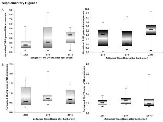
Download
1 / 1
10 likes | 172 Views
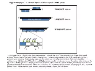
Supplementary figure 1. A schematic figure of the three-segmented RVFV genome.

E N D
Supplementary figure 1. A schematic figure of the three-segmented RVFV genome Supplementary figure 1 illustrates the three-segmented RVFV genome, the size of the three RNA segments and the encoded proteins. The upper part of the figure show the S segment and the two genes encoding the N and NSs protein with the inter-genomic region separating the two coding sequences. The middle part of this figure demonstrate the L segment and the multifunctional RNA dependent RNA polymerase (RdRp) protein encoded in an antisense manner. The cartoon at the bottom of the figure shows the M segment and the polyprotein precursor that is subsequently cleaved into the NSm, Gn and Gc proteins. The five potential in frame translation initiation codons are shown below in a magnified picture of the NSm encoding region. The F and the R primers used to amplify the NSm gene, from the proposed second AUG codon, are also shown.