Download

1 / 35
350 likes | 762 Views
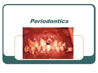
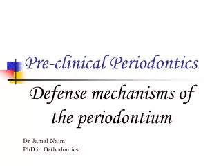
Pre-clinical Periodontics. Dr Jamal Naim PhD in Orthodontics. Introduction cont. Definitions. Carranza's - clinical periodontology. Cementum. Cemento-enamel junction (CEJ) Cementicles Hypercementosis Root resporption Ankylosis Exposure of cementum. Cementoenamel junction.

E N D
Pre-clinical Periodontics Dr Jamal Naim PhD in Orthodontics Introduction cont.
Definitions Carranza's - clinical periodontology
Cementum • Cemento-enamel junction (CEJ) • Cementicles • Hypercementosis • Root resporption • Ankylosis • Exposure of cementum
Cementoenamel junction The relation between, cementum and enamel at the cervical region of teeth is variable. • In approximately 60% of the teeth, cementum overlaps the cervical end of enamel for a short distance (1). • In approximately 30% of all teeth, cementum meets the cervical end of enamel in a relatively sharp line (2). 1 2 3
Cementoenamel junction In 10% of the cases there is no cementoenamel junction. Instead, a zone of the root is devoid of cementum and is, for a time, covered by reduced enamel epithelium. Gingival recession may result by such cases more sensitivity to irritants due to dentine exposure.
Cementicles Cementicles are small-mineralized bodies, which may be found in the periodontal ligament. They may be attached to the cementum or the alveolar bone, or occur free in the periodontal ligament. When present, cementicles are generally found about all or most of the teeth. Cementicles may be formed by mineralization of degenerating epithelial rests or thrombosed vessels.
Hypercementosis Hypercementosis: It is an abnormal thickening of cementum, may be diffuse or circumscribed. It may affect all teeth of the dentition, be confined to a single tooth, or even affect only Parts of one tooth. If the overgrowth improves the functional qualities of the cementum, it is termed a cementum hypertrophy. If the overgrowth occurs in nonfunctional teeth, it is termed hyperplasia (e.g. ostitis deformans paget).
Localized hypercementosis Generalized hypercementosis
Hypercementosis In Localized hypertrophy prong like extension of cementum may be formed. This condition frequently is found in teeth that are exposed to great stress (ortho) (compensatory cementum). This extension of cementum provide a larger surface area for the attaching fibers; thus a firmer anchorage of the tooth to the surrounding alveolar bone is assured. Localized hypercementosis may some times due periapical inflammation. Here the hyperplasia is circumscribed and surrounds the root like a cuff.
Root resorption Although root resorption is not a part of the normal functional activity in the permanent dentition, most teeth show minute areas of resorption which may extend through the cementum and into the root dentin. RR is painless. Causes of root resorption: • Trauma from occlusion, orthodontic forces or accidents • Pressure from erupting teeth, cysts or tumors • Periapical or periodontal diseases • Teeth without function and transplanted teeth • unknown reasons (idiopathic).
Root resorption Some systemic conditions induce root resorption: • Calcium deficiency • Hypothyroidism • Fibrous osteodystrophy • Pagets disease The defect is repaired by rapid deposition of cellular cementum once the initiating factor is removed (not by mentioned systemic conditions)
Ankylosis Ankylosis is the fusion of cementum and alveolar boe with obliteration of the periodontal ligament. It may be a form of abnormal repair of root resorption. Ankylosis results mostly in root resorption and replacement by bone tissue.
Exposure of cementum By gingival recessions and attachment loss cementum will be exposed to oral environment. • Cementum is permeable for organic and inorganic substances • Bacterial invasion of cementum can cause root caries
Definitions Carranza's - clinical periodontology
Alveolar bone The alveolar process is that bone containing the alveoli. It consists of: • an outer (lingual and buccal) cortical plate (compact bone) • A central spongiosa (spongous bone) and • Alveolar bone proper (bone lining the alveolus), (bundle bone) • The alveolar bone and the cortical plate meet at the alveolar crest (1.5 to 2 mm below the level of CEJ).
Alveolar bone proper • It surrounds the roots of the tooth & gives attachment to the principal fibers of the periodontal ligament. • It also known as the cribiform plate as it is perforated by many openings; through which branches of the interalveolar nerve & vessels pass into the periodontal ligament. • The bone which lines the socket in which sharpey’s fibers are embedded is known as bundle bone. • Contains more calcium salts per units area than other bone. • It also know as lamina dura because of its radiopacity.
Alveolar bone proper In X-ray the cribriform plate is referred to lamina dura. Cribriform plate
Alveolar bone proper • Massive cortical plate • thin lamina dura • In between trabecular bone
The cortical plates The cortical plate of alveolar process consists of fine fibered lamellar bone. It is generally thinner in the maxilla and thickest on the buccal aspect of the mandibular molars and premolars.
The cortical plates Trabecular bone is absent in the region of frontal teeth, so that cortical plate and alveolar bone are fused together.
Osseous topography Normally: prominence of the roots with the intervening vertical depressions that taper toward the margin. On the labial version; the margins of the labial bone is thinned to a knife edge & presents an apically accentuated arc.
Osseous topography On the lingual version ; the margins of the labial bone is blunt & rounded & horizontal rather than arcuate.
FENESTRATIONS & DEHISCENCES • Isolated areas in which the root is denuded of bone & the root surface is covered only by periosteum & overlying gingiva are termed fenestrations. • When the denuded areas extends through the marginal bone then defect is called a dehiscence.
BONE TURNOVER (REMODELLING) • The process by which the overall size & shape of bones is established is referred to as bone remodeling. • Alveolar bone builds a stable cast for the periodontal tissues because its structure is in a constant state of flux. Influencing factors Local: • Functional requirements on the tooth • Age related changes in the bone cells Systemic: • Parathyroid hormone • Calcitonin • Vitamin D3
Physiologic tooth mobility • Each tooth has a physiologic mobility in horizontal vertical and rotational direction • It varies among healthy persons • It varies also within 24hour cycle, the teeth are more mobile in the morning than in the evening • The mobility depends on Root surface area available for the insertion of sharpey's fibers: • Number of roots • Length of roots • Diameter of roots
Physiologic tooth mobility Mobility of various tooth types
Physiologic tooth mobility • Elevated tooth mobility is not a criterion for periodontal health or pathology • The mobility increase by trauma and inflammation
Initial tooth mobility • It is the first phase of movement after application of 100g force in labiolingual direction • The tooth moves easily in the socket • There is no bony deformation • Some periodontal fiber bundles are stretched others are relaxed • It depends on the width of the PDL and the histology of it • It ranges from 5/100 to 10/100 mm
Secondary tooth mobility • It is measured after application of 500g force in labiolingual direction • The tooth moves easily in the socket then causes bony deformation of he entire alveolar process • The magnitude of deformation depends on: • Quality • Thickness • Elasticity of the alveolar bone • It ranges from 8/100 to 15/100 mm
Physiologic tooth mobility g g Initial and secondary tooth Mobility
Characteristics Associated with Clinical Gingival Health • Absence of inflammation • No exudate • No bleeding on probing (BOP)
Characteristics Associated with Histological Gingival Health Most people have histological signs of gingivitis but may not be seen clinically