Download
1 / 29
290 likes | 578 Views
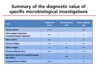
Summary of the diagnostic value of specific microbiological investigations. Blood culture. Technique Minimal 1~3 mL each Different venipuncture ( ≥ 2 sets) Central catheter + Peripheral culture Colonized bacteria – possibility of contamination. Blood culture. Blood culture. Gram stain.

E N D
Summary of the diagnostic value of specific microbiological investigations
Blood culture • Technique • Minimal 1~3 mL each • Different venipuncture(≥2 sets) • Central catheter + Peripheral culture • Colonized bacteria –possibility of contamination
Blood culture Gram stain
Interpretation of blood culture • Possible contaminant • P. acnes • Corynebacteriumspecies • Bacillus species • CoNS (coagulase negative Staphylococci, S. epidermidis) • S. viridans • Peptostreptococcus Most causative pathogens are compatible with clinical syndrome Most causative pathogens will grow within 72 hours • Almost always important • S. aureus • S. pneumoniae • S. pyogenes(GAS) • S. agalactiae(GBS) • H. influenzae • Enterobacteriaceae(E. coli, Klebsiella, etc..) • Bacteroidaceae • P. aeruginosa • Candida species
Nasopharyngeal aspirate collection • Nasopharyngeal aspirate – Virus culture / PCR
Early diagnosis of viral infection • Microscopy • LM • EM • Detecting antigen • IF staining • Solid phase immunoassay (ELISA, RIA, LA), • Immunochromatography • Detecting nucleic acid • nucleic acid amplification (PCR, etc.), • nucleic acid hybridization
Ultrastructural Characteristics of SARS-Associated Coronavirus Grown in Vero E6 Cell 100 nm
Indirect IF Staining for RSV, • Specimen: Nasal aspirate
Indirect IF Staining for PIV3 • Specimen: Nasal aspirate • Confocal microscopy
Direct Method Antigen Detection by ELISA Indirect Method
Indirect ELISA for RSV Specimen: Nasal aspirate
Conventional method vs.RT-PCR Virus identified Detection by conventional method (% of total episodes) No. detected by RT-PCR (% of total episodes) Respiratory syncytial virus 122 (23.7) 596 (14.7) Adenovirus 35 (6.8) 243 (6.0) Parainfluenza virus type1 11 (2.1) 30 (0.7) type 2 ND 23 (0.6) type 3 32 (6.2) 195 (4.8) type 4 ND 2 (0.05) Influenza virus A 24 (4.7) 117 (2.9) B 9 (1.8) 47 (1.2) ND ND ND ND ND 17 (0.3) Rhinovirus Metapneumovirus Coronavirus, conventional Coronavirus NL63 Bocavirus Unknown 40 (7.8) 26 (5.0) 1 (<1) 9 (1.8) 58 (11.2) Total 316 (61.4) * 1,270 (30.4) ** *More than 1 virus were isolated from 36 patients (11.4%) among 514 patients, Sep 2000-Aug 2005. **1,270 strains of viruses were isolated from 1,234 out of 4,058 cases (30.4% of total patients), Nov 1990-May 2002. More than 1 virus were isolated from 36 patients. Choi EH et al. Clin Infect Dis 2006;43:585-92