Download
1 / 15
220 likes | 573 Views
Mechanisms of Gene Mutation. Lecture 7 Dr. Attya Bhatti. Molecular basis of gene mutations. Are of two types 1. Spontaneous mutations 2. Induced mutations Can be analyzed by Molecular Genetic Techniques. Spontaneous mutations. Arise from a variety of sources,

E N D
Mechanisms of Gene Mutation Lecture 7 Dr. AttyaBhatti
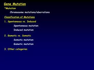
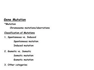
Molecular basis of gene mutations • Are of two types 1. Spontaneous mutations 2. Induced mutations • Can be analyzed by Molecular Genetic Techniques.
Spontaneous mutations • Arise from a variety of sources, • Errors in DNA replication, • Spontaneous lesions, • Transposable genetic elements.
Errors in DNA Replication Pairing between the normal (keto) forms of the bases.
Errors in DNA Replication Mismatched bases. (a) Mispairs resulting from rare tautomeric forms of the pyrimidines; (b) mispairs resulting from rare tautomeric forms of the purines. A keto structure occurs when the hydrogen atom bonds to a nitrogen atom within the ring. An enol structure occurs when the hydrogen atom bonds to an nearby oxygen atom that sticks out from the ring. These two types of structures are known as tautomers.
Errors in DNA Replication • Results in • Transitions • Transversions • Frameshift mutations • Deletions and duplications
Spontaneous lesions • Result from depurination and deamination. • Depurination, the more common of the two, consists of the interruption of the glycosidic bond between the nitrogenous base and deoxyribose and the subsequent loss of a guanine or an adenine residue from the DNA. Fig: The loss of a purine residue (guanine) from a single strand of DNA. The sugar-phosphate backbone is left intact.
Spontaneous lesions • The deamination of cytosine yields uracil. • Unrepaired uracil residues will pair with adenine in replication, resulting in the conversion of a G–C pair into an A–T pair (a GC → AT transition). Fig: Deamination of (a) cytosine and (b) 5-methylcytosine.
Spontaneous mutations and human diseases • Disorders are due to deletions or duplications involving repeated sequences. • For example, mitochondrial encephalomyopathies are a group of disorders affecting the central nervous system or the muscles (Kearns-Sayre syndrome). Fig: Sequences of wild-type (WT) mitochondrial DNA and deleted DNA (KS) from a patient with Kearns-Sayre syndrome. The 13-base boxed sequence is identical in both WT and KS and serves as a breakpoint for the DNA deletion. A single base (boldface type) is altered in KS, aside from the deleted segment.
Spontaneous mutations and human diseases • Genetic diseases due to the expansion of a three-base-pair repeat. • For example; Fragile X syndrome Fig: Expansion of the CGG triplet in the FMR-1 gene seen in the fragile X syndrome. Normal persons have from 6 to 54 copies of the CGG repeat, whereas members of susceptible families display an increase (premutation) in the number of repeats: normally transmitting males (NTMs) and their daughters are phenotypically normal but display from 50 to 200 copies of the CGG triplet; the number of repeats expands to some 200 to 1300 in those showing full symptoms of the disease.
Induced mutations • Introduction of mutations by mutagens. • Mutagens induce mutations by at least three different mechanisms. • Incorporation of base analogs. • Specific mispairing. • Base damage.
Incorporation of base analogs Fig: Alternative pairing possibilities for 5-bromouracil (5-BU). 5-BU is an analog of thymine that can be mistakenly incorporated into DNA as a base. It has a bromine atom in place of the methyl group. (a) In its normal keto state, 5-BU mimics the pairing behavior of the thymine that it replaces, pairing with adenine. (b) The presence of the bromine atom, however, causes a relatively frequent redistribution of electrons, so that 5-BU can spend part of its existence in the rare ionized form. In this state, it pairs with guanine, mimicking the behavior of cytosine and thus inducing mutations in replication.
Specific mispairing • Some mutagens are not incorporated into the DNA but instead alter a base, causing specific mispairing. • E.g Certain alkylating agents, such as ethylmethanesulfonate (EMS) and Intercalating agents such as proflavin, acridine orange. Fig: Intercalating agents. (a) Structures of the common agents proflavin, acridine orange, and ICR-191. (b) An intercalating agent slips between the nitrogenous bases stacked at the center of the DNA molecule. This occurrence can lead to single-nucleotide-pair insertions and deletions.
Base damage • A large number of mutagens damage one or more bases, so no specific base pairing is possible. • The result is a replication block, because DNA synthesis will not proceed past a base that cannot specify its complementary partner by hydrogen bonding.
Base damage • Ionizing radiation can cause breakage of the N-glycosydic bond, leading to the formation of AP sites, and can cause strand breaks that are responsible for most of the lethal effects of such radiation. • AP site • Apurinic or apyrimidinic site resulting from the loss of a purine or pyrimidine residue from the DNA