Download
1 / 28
521 likes | 3.28k Views
Mechanisms of Gene Mutation. Lecture 7 Dr. Attya Bhatti. Molecular basis of gene mutations. Are of two types 1. Spontaneous mutations 2. Induced mutations Can be analyzed by Molecular Genetic Techniques. Spontaneous mutations. Arise from a variety of sources,

E N D
Mechanisms of Gene Mutation Lecture 7 Dr. AttyaBhatti
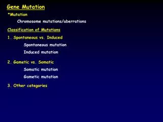
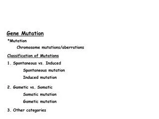
Molecular basis of gene mutations • Are of two types 1. Spontaneous mutations 2. Induced mutations • Can be analyzed by Molecular Genetic Techniques.
Spontaneous mutations • Arise from a variety of sources, • Errors in DNA replication, • Spontaneous lesions, • Transposable genetic elements.
Errors in DNA Replication Pairing between the normal (keto) forms of the bases.
Errors in DNA Replication Mismatched bases. (a) Mispairs resulting from rare tautomeric forms of the pyrimidines; (b) mispairs resulting from rare tautomeric forms of the purines. A keto structure occurs when the hydrogen atom bonds to a nitrogen atom within the ring. An enol structure occurs when the hydrogen atom bonds to an nearby oxygen atom that sticks out from the ring. These two types of structures are known as tautomers.
Errors in DNA Replication • Results in • Transitions • Transversions • Frameshift mutations • Deletions and duplications
Spontaneous lesions • Result from depurination and deamination. • Depurination, the more common of the two, consists of the interruption of the glycosidic bond between the nitrogenous base and deoxyribose and the subsequent loss of a guanine or an adenine residue from the DNA. Fig: The loss of a purine residue (guanine) from a single strand of DNA. The sugar-phosphate backbone is left intact.
Spontaneous lesions • The deamination of cytosine yields uracil. • Unrepaired uracil residues will pair with adenine in replication, resulting in the conversion of a G–C pair into an A–T pair (a GC → AT transition). Fig: Deamination of (a) cytosine and (b) 5-methylcytosine.
Spontaneous mutations and human diseases • Disorders are due to deletions or duplications involving repeated sequences. • For example, mitochondrial encephalomyopathies are a group of disorders affecting the central nervous system or the muscles (Kearns-Sayre syndrome). Fig: Sequences of wild-type (WT) mitochondrial DNA and deleted DNA (KS) from a patient with Kearns-Sayre syndrome. The 13-base boxed sequence is identical in both WT and KS and serves as a breakpoint for the DNA deletion. A single base (boldface type) is altered in KS, aside from the deleted segment.
Spontaneous mutations and human diseases • Genetic diseases due to the expansion of a three-base-pair repeat. • For example; Fragile X syndrome Fig: Expansion of the CGG triplet in the FMR-1 gene seen in the fragile X syndrome. Normal persons have from 6 to 54 copies of the CGG repeat, whereas members of susceptible families display an increase (premutation) in the number of repeats: normally transmitting males (NTMs) and their daughters are phenotypically normal but display from 50 to 200 copies of the CGG triplet; the number of repeats expands to some 200 to 1300 in those showing full symptoms of the disease.
Induced mutations • Introduction of mutations by mutagens. • Mutagens induce mutations by at least three different mechanisms. • Incorporation of base analogs. • Specific mispairing. • Base damage.
Incorporation of base analogs Fig: Alternative pairing possibilities for 5-bromouracil (5-BU). 5-BU is an analog of thymine that can be mistakenly incorporated into DNA as a base. It has a bromine atom in place of the methyl group. (a) In its normal keto state, 5-BU mimics the pairing behavior of the thymine that it replaces, pairing with adenine. (b) The presence of the bromine atom, however, causes a relatively frequent redistribution of electrons, so that 5-BU can spend part of its existence in the rare ionized form. In this state, it pairs with guanine, mimicking the behavior of cytosine and thus inducing mutations in replication.
Specific mispairing • Some mutagens are not incorporated into the DNA but instead alter a base, causing specific mispairing. • E.g Certain alkylating agents, such as ethylmethanesulfonate (EMS) and Intercalating agents such as proflavin, acridine orange. Fig: Intercalating agents. (a) Structures of the common agents proflavin, acridine orange, and ICR-191. (b) An intercalating agent slips between the nitrogenous bases stacked at the center of the DNA molecule. This occurrence can lead to single-nucleotide-pair insertions and deletions.
Base damage • A large number of mutagens damage one or more bases, so no specific base pairing is possible. • The result is a replication block, because DNA synthesis will not proceed past a base that cannot specify its complementary partner by hydrogen bonding.
Base damage • Ionizing radiation can cause breakage of the N-glycosydic bond, leading to the formation of AP sites, and can cause strand breaks that are responsible for most of the lethal effects of such radiation. • AP site • Apurinic or apyrimidinic site resulting from the loss of a purine or pyrimidine residue from the DNA
Relation between mutagens and carcinogens • 157 of 175 known carcinogens (approximately 90 percent) are mutagens. Induced mutations and human cancer • Ultraviolet light and aflatoxin B1have long been suspected of causing skin cancer and liver cancer, respectively. • DNA sequence analysis of mutations in a human cancer gene has provided direct evidence of their involvement. • p53 is one of a number of tumor-suppressor genes that encode proteins that suppress tumor formation. • A sizable proportion of human cancer patients have mutatedtumor-suppressor genes.
Biological repair mechanisms • Living cells have evolved a series of enzymatic systems that repair DNA damage in a variety of ways. • Prevention of errors before they happen • Direct reversal of damage • Excision-repair pathways • Post replication repair
Prevention of errors before they happen • Some enzymatic systems neutralize potentially damaging compounds before they even react with DNA. • One example of such a system is the detoxification of superoxide radicals produced during oxidative damage to DNA: the enzyme superoxide dismutase catalyzes the conversion of the superoxide radicals into hydrogen peroxide, and the enzyme catalase, in turn, converts the hydrogen peroxide into water.
Direct reversal of damage Fig: Repair of a UV-induced pyrimidine photodimer by a photoreactivating enzyme, or photolyase. The enzyme recognizes the photodimer (here, a thymine dimer) and binds to it. When light is present, the photolyase uses its energy to split the dimer into the original monomers.
Excision-repair pathways • In E. coli, the products of the uvrA, B, and C genes constitute the excinuclease. • The UvrA protein, which recognizes the damaged DNA, forms a complex with UvrB and leads the UvrB subunit to the damage site before dissociating. • The UvrC protein then binds to UvrB. Each of these subunits makes an incision. • The short DNA 12-mer is unwound and released by another protein, helicase II. • The human excinuclease is considerably more complex than its bacterial counterpart and includes at least 17 proteins. However, the basic steps are the same as those in E. coli.
Excision-repair pathways Fig: Schematic representation of events following incision by UvrABC exinuclease in E. coli. First, the UvrA subunit recruits the UvrB subunit to the damage site before dissociation. Then, as shown here, DNA helicase II mediates the release of a segment of the DNA bounded by two nicks in the same strand of DNA. The UvrC protein is also displaced at this point. The subsequent repair synthesis displaces UvrB.
Coupling of transcription and repair • In both eukaryotes and prokaryotes, there is a preferential repair of the transcribed strand of DNA for actively expressed genes. Fig: Nucleotide excision repair is coupled to transcription. This model for coupled repair in mammalian cells shows RNA polymerase (pink) pausing when encountering a lesion. It undergoes a conformational change, allowing the DNA strands at the lesion site to reanneal. Protein factors aid in coupling by bringing TFIIH and other factors to the site to carry out the incision, excision, and repair reactions. Then transcription can continue normally.
Postreplication repair • Mismatch repair • Recombinational repair • Mismatch repair • Some repair pathways are capable of recognizing errors even after DNA replication has already occurred. • One such system, termed the mismatch repair system, can detect mismatches that occur in DNA replication.
Mismatch repair • Works in three steps; • Recognize mismatched base pairs. • Determine which base in the mismatch is the incorrect one. • Excise the incorrect base and carry out repair synthesis.
Mismatch repair in humans. (1) Mispairs and misaligned bases arise in the course of replication. (2) The G–T-binding protein (GTBP) and the human MutS homolog (hMSH2) recognize the incorrect matches. (3) Two additional proteins, hPMS2 and hMLH1, are recruited and form a larger repair complex. (4) The mismatch is repaired after removal, DNA synthesis, and ligation.
Recombinational repair Schemes for postreplication repair. (a) In recombinational repair, replication jumps across a blocking lesion, leaving a gap in the new strand. A recA-directed protein then fills the gap, using a piece from the opposite parental strand (because of DNA complemen-tarity, this filler will supply the correct bases for the gap). Finally, the RecA protein repairs the gap in the parental strand.
Repair defects and human diseases Table: Human Diseases with DNA-Repair Defects The production of a wild-type phenotype when two different mutations are combined in a diploid or a heterokaryon called complementation.
Assignment: Chromosomal aberrations Topic 1: Changes in the number of chromosomes. Aneuploidy and euploidy. Topic 2: Changes in the structure of chromosomes, deficiency, duplication, inversion and translocation. Marks distribution: Data collection, arrangement of data, Comprehensive description of topic. How well described? Group: Maximum three students in a group. Explain contribution of each student in one line.